The Cellular Level of Organization
3.0 Introduction
Chapter Targets
After learning this chapter, it is possible for you to to:
- Describe the construction and performance of the cell membrane, together with its regulation of supplies into and out of the cell
- Describe the features of the varied cytoplasmic organelles
- Listing the morphological and physiological traits of some consultant cell sorts within the human physique
- Clarify the construction and contents of the nucleus, in addition to the method of DNA replication
- Clarify the method by which a cell builds proteins utilizing the DNA code
- Listing the levels of the cell cycle so as, together with the steps of cell division in somatic cells
- Focus on how a cell differentiates and turns into extra specialised
You developed from a single fertilized egg cell into the complicated organism that you simply see whenever you look in a mirror, containing trillions of cells. Throughout this developmental course of, early, unspecialized cells turn into specialised of their construction and performance (this is named differentiation). These completely different cell sorts be part of to type specialised tissues that work in live performance to carry out the entire features needed for the residing organism. Mobile and developmental biologists research how the continued division of a single cell results in such complexity and differentiation.
Contemplate the distinction between a structural cell within the pores and skin and a nerve cell. A structural pores and skin cell could also be formed like a flat plate (squamous) and stay just for a short while earlier than it’s shed and changed. Packed tightly into rows and sheets, the squamous pores and skin cells present a protecting barrier for the cells and tissues that lie beneath. A nerve cell, however, could also be formed one thing like a star, sending out lengthy processes as much as a meter in size and should stay for the whole lifetime of the organism. With their lengthy winding appendages, nerve cells can talk with each other and with different forms of physique cells and ship speedy alerts that inform the organism about its setting and permit it to work together with that setting. These variations illustrate one crucial theme that’s constant in any respect organizational ranges of biology: the type of a construction is optimally suited to carry out explicit features assigned to that construction. Hold this theme in thoughts as you tour the within of a cell and are launched to the varied forms of cells within the physique.
A main accountability of every cell is to contribute to homeostasis. Homeostasis is a time period utilized in biology that refers to a dynamic state of steadiness inside parameters which might be appropriate with life. For instance, residing cells require a water-based setting to outlive in, and there are numerous bodily (anatomical) and physiological mechanisms that hold the entire trillions of residing cells within the human physique moist. That is one facet of homeostasis. When a selected parameter, comparable to blood strain or blood oxygen content material, strikes far sufficient out of homeostasis (usually turning into too excessive or too low), sickness or illness—and generally dying—inevitably outcomes.
The idea of a cell began with microscopic observations of useless cork tissue by scientist Robert Hooke in 1665. With out realizing their operate or significance, Hooke coined the time period “cell” primarily based on the resemblance of the small subdivisions within the cork to the rooms that monks inhabited, referred to as cells. About ten years later, Antonie van Leeuwenhoek grew to become the primary particular person to look at residing and transferring cells underneath a microscope. Within the century that adopted, the idea that cells represented the fundamental unit of life would develop. These tiny fluid-filled sacs home parts accountable for the 1000’s of biochemical reactions needed for an organism to develop and survive. On this chapter, you’ll be taught concerning the main parts and features of cells and uncover among the various kinds of cells within the human physique.
3.1 The Cell Membrane
Studying Targets
By the top of this part, it is possible for you to to:
- Describe the molecular parts that make up the cell membrane
- Relate buildings of the cell membrane to its features
- Describe how molecules cross the cell membrane primarily based on their properties and focus gradients
- Examine and distinction various kinds of passive transport with lively transport, offering examples of every
Regardless of variations in construction and performance, all residing cells in multicellular organisms have a surrounding cell membrane. Simply because the outer layer of your pores and skin separates your physique from its setting, the cell membrane (also referred to as the plasma membrane) separates the inside contents of a cell from its exterior setting. This cell membrane offers a protecting barrier across the cell and regulates which supplies can move in or out.
Construction and Composition of the Cell Membrane
The cell membrane is an especially pliable construction composed primarily of two layers of phospholipids (a “bilayer”). Ldl cholesterol and varied proteins are additionally embedded throughout the membrane giving the membrane a wide range of features described under.
A single phospholipid molecule has a phosphate group on one finish, referred to as the “head,” and two side-by-side chains of fatty acids that make up the lipid “tails” (Determine 3.1.1). The lipid tails of 1 layer face the lipid tails of the opposite layer, assembly on the interface of the 2 layers. The phospholipid heads face outward, one layer uncovered to the inside of the cell and one layer uncovered to the outside (Determine 3.1.1).
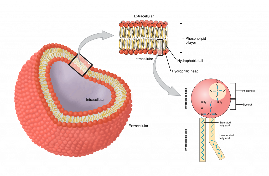
The phosphate group is negatively charged, making the top polar and hydrophilic—or “water loving.” A hydrophilic molecule (or area of a molecule) is one that’s interested in water. The phosphate heads are thus interested in the water molecules of each the extracellular and intracellular environments. The lipid tails, however, are uncharged, or nonpolar, and are hydrophobic—or “water fearing.” A hydrophobic molecule (or area of a molecule) repels and is repelled by water. Phospholipids are thus amphipathic molecules. An amphipathic molecule is one which incorporates each a hydrophilic and a hydrophobic area. In reality, cleaning soap works to take away oil and grease stains as a result of it has amphipathic properties. The hydrophilic portion can dissolve within the wash water whereas the hydrophobic portion can entice grease in stains that then could be washed away. An analogous course of happens in your digestive system when bile salts (constructed from ldl cholesterol, phospholipids and salt) assist to interrupt up ingested lipids.
Because the phosphate teams are polar and hydrophilic, they’re interested in water within the intracellular fluid. Intracellular fluid (ICF) is the fluid inside of the cell. The phosphate teams are additionally interested in the extracellular fluid. Extracellular fluid (ECF) is the fluid setting exterior the enclosure of the cell membrane (see above Determine). Because the lipid tails are hydrophobic, they meet within the inside area of the membrane, excluding watery intracellular and extracellular fluid from this area. Along with phospholipids and ldl cholesterol, the cell membrane has many proteins detailed within the subsequent part.
Membrane Proteins
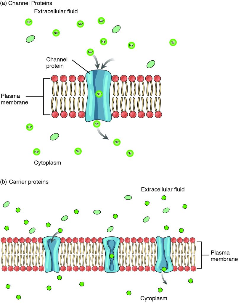
The lipid bilayer kinds the premise of the cell membrane, however it’s peppered all through with varied proteins. Two various kinds of proteins which might be generally related to the cell membrane are the integral protein and peripheral protein (Determine 3.1.2). As its title suggests, an integral protein is a protein that’s embedded within the membrane. Many various kinds of integral proteins exist, every with completely different features. For instance, an integral protein that extends a gap by way of the membrane for ions to enter or exit the cell is named a channel protein. Peripheral proteins are sometimes discovered on the inside or outer floor of the lipid bilayer however can be connected to the inner or exterior floor of an integral protein.
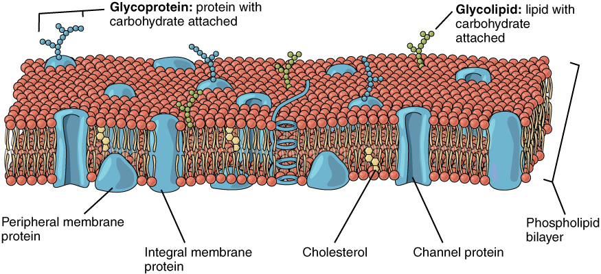
Some integral proteins function cell recognition or floor id proteins, which mark a cell’s id in order that it may be acknowledged by different cells. Some integral proteins act as enzymes, or in cell adhesion, between neighboring cells. A receptor is a sort of recognition protein that may selectively bind a particular molecule exterior the cell, and this binding induces a chemical response throughout the cell. Some integral proteins serve twin roles as each a receptor and an ion channel. One instance of a receptor-channel interplay is the receptors on nerve cells that bind neurotransmitters, comparable to dopamine. When a dopamine molecule binds to a dopamine receptor protein, a channel throughout the transmembrane protein opens to permit sure ions to move into the cell. Peripheral proteins are sometimes related to integral proteins alongside the inside cell membrane the place they play a task in cell signaling or anchoring to inner mobile parts (ie: cytoskeleton mentioned later).
Some integral membrane proteins are glycoproteins. A glycoprotein is a protein that has carbohydrate molecules connected, which lengthen into the extracellular setting. The connected carbohydrate tags on glycoproteins help in cell recognition. The carbohydrates that stretch from membrane proteins and even from some membrane lipids collectively type the glycocalyx. The glycocalyx is a fuzzy-appearing coating across the cell shaped from glycoproteins and different carbohydrates connected to the cell membrane. The glycocalyx can have varied roles. For instance, it might have molecules that permit the cell to bind to a different cell, it might comprise receptors for hormones, or it might need enzymes to interrupt down vitamins. The glycocalyces present in an individual’s physique are merchandise of that particular person’s genetic make-up. They provide every of the person’s trillions of cells the “id” of belonging within the particular person’s physique. This id is the first means that an individual’s immune protection cells “know” to not assault the particular person’s personal physique cells, but it surely is also the rationale organs donated by one other particular person could be rejected.
Transport Throughout the Cell Membrane
One of many nice wonders of the cell membrane is its potential to control the focus of gear contained in the cell. These substances embrace ions comparable to Ca++, Na+, Okay+, and Cl–, vitamins together with sugars, fatty acids, and amino acids, and waste merchandise, significantly carbon dioxide (CO2), which should depart the cell.
The membrane’s lipid bilayer construction offers the primary degree of management. The phospholipids are tightly packed collectively, and the membrane has a hydrophobic inside. This construction causes the membrane to be selectively permeable. A membrane that has selective permeability permits solely substances assembly sure standards to move by way of it unaided. Within the case of the cell membrane, solely comparatively small, nonpolar supplies can transfer by way of the lipid bilayer (bear in mind, the lipid tails of the membrane are nonpolar). Some examples of those are different lipids, oxygen and carbon dioxide gases, and alcohol. Nonetheless, water-soluble supplies—like glucose, amino acids, and electrolytes—want some help to cross the membrane as a result of they’re repelled by the hydrophobic tails of the phospholipid bilayer. All substances that transfer by way of the membrane accomplish that by considered one of two common strategies, that are categorized primarily based on whether or not or not power is required. Passive transport is the motion of gear throughout the membrane with out the expenditure of mobile power. In distinction, lively transport is the motion of gear throughout the membrane utilizing power from adenosine triphosphate (ATP).
Passive Transport
With a purpose to perceive how substances transfer passively throughout a cell membrane, it’s needed to grasp focus gradients and diffusion. A focus gradient is the distinction in focus of a substance throughout an area. Molecules (or ions) will unfold/diffuse from the place they’re extra concentrated to the place they’re much less concentrated till they’re equally distributed in that area. (When molecules transfer on this means, they’re mentioned to maneuver down their focus gradient, from excessive focus to low focus.) Diffusion is the motion of particles from an space of upper focus to an space of decrease focus. A few widespread examples will assist as an instance this idea. Think about being inside a closed room. If a bottle of fragrance had been sprayed, the scent molecules would naturally diffuse from the spot the place they left the bottle to all corners of the room, and this diffusion would go on till the molecules had been equally distributed within the room. One other instance is a spoonful of sugar positioned in a cup of tea. Ultimately the sugar will diffuse all through the tea till no focus gradient stays. In each instances, if the room is hotter or the tea hotter, diffusion happens even sooner because the molecules are bumping into one another and spreading out sooner than at cooler temperatures.
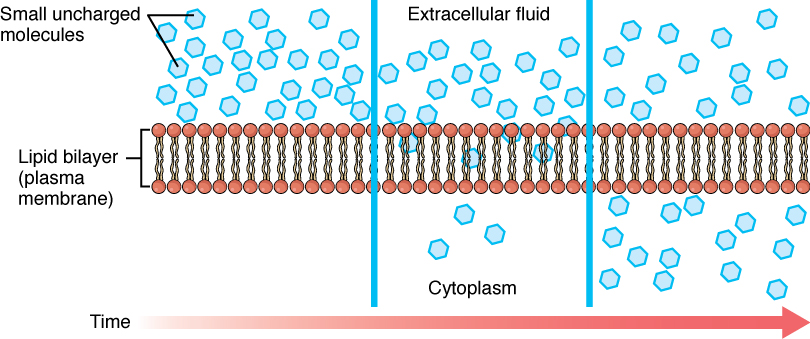
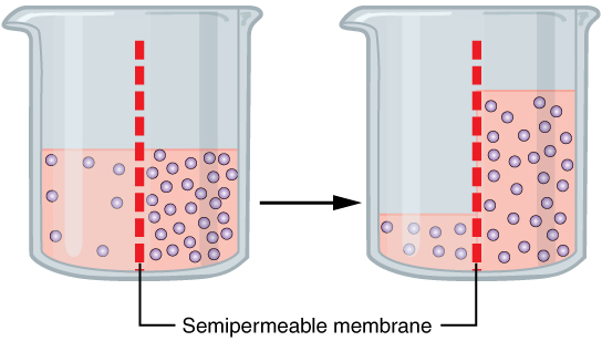
Every time a substance exists in higher focus on one aspect of a semipermeable membrane, comparable to cell membranes, any substance that may transfer down its focus gradient throughout the membrane will accomplish that. If the substances can transfer throughout the cell membrane with out the cell expending power, the motion of molecules known as passive transport. Contemplate substances that may simply diffuse by way of the lipid bilayer of the cell membrane, such because the gases oxygen (O2) and carbon dioxide (CO2). These small, fats soluble gasses and different small lipid soluble molecules can dissolve within the membrane and enter or exit the cell following their focus gradient. This mechanism of molecules transferring throughout a cell membrane from the aspect the place they’re extra concentrated to the aspect the place they’re much less concentrated is a type of passive transport referred to as easy diffusion. O2 usually diffuses into cells as a result of it’s extra concentrated exterior of them, and CO2 sometimes diffuses out of cells as a result of it’s extra concentrated within them.
Earlier than transferring on, you will need to notice that the focus gradients for oxygen and carbon dioxide will all the time exist throughout a residing cell and by no means attain equal distribution. It is because cells quickly expend oxygen throughout metabolism and so, there’s sometimes a decrease focus of O2 contained in the cell than exterior. In consequence, oxygen will diffuse from exterior the cell instantly by way of the lipid bilayer of the membrane and into the cytoplasm throughout the cell. Alternatively, as a result of cells produce CO2 as a byproduct of metabolism, CO2 concentrations rise throughout the cytoplasm; subsequently, CO2 will transfer from the cell by way of the lipid bilayer and into the extracellular fluid, the place its focus is decrease. (Determine 3.1.3).
Massive polar or ionic molecules, that are hydrophilic, can’t simply cross the phospholipid bilayer. Charged atoms or molecules of any dimension can’t cross the cell membrane by way of easy diffusion as the fees are repelled by the hydrophobic tails within the inside of the phospholipid bilayer. Solutes dissolved in water on both aspect of the cell membrane will are inclined to diffuse down their focus gradients, however as a result of most substances can’t move freely by way of the lipid bilayer of the cell membrane, their motion is restricted to protein channels and specialised transport mechanisms within the membrane. Facilitated diffusion is the diffusion course of used for these substances that can’t cross the lipid bilayer resulting from their dimension, cost, and/or polarity however accomplish that down their focus gradients (Determine 3.1.4). For example, despite the fact that sodium ions (Na+) are extremely concentrated exterior of cells, these electrolytes are charged and can’t move by way of the nonpolar lipid bilayer of the membrane. Their diffusion is facilitated by membrane proteins that type sodium channels (or “pores”), in order that Na+ ions can transfer down their focus gradient from exterior the cells to contained in the cells. A typical instance of facilitated diffusion utilizing a provider protein is the motion of glucose into the cell, the place it’s used to make ATP. Though glucose could be extra concentrated exterior of a cell, it can’t cross the lipid bilayer by way of easy diffusion as a result of it’s each giant and polar, and subsequently, repelled by the phospholipid membrane. To resolve this, a specialised provider protein referred to as the glucose transporter will switch glucose molecules into the cell to facilitate its inward diffusion. The distinction between a channel and a provider is that the provider normally adjustments form in the course of the diffusion course of, whereas the channel doesn’t. There are lots of different solutes that should bear facilitated diffusion to maneuver right into a cell, comparable to amino acids, or to maneuver out of a cell, comparable to wastes.
Osmosis
A specialised instance of facilitated transport is water transferring throughout the cell membrane of all cells, by way of protein channels generally known as aquaporins. Osmosis is the diffusion of water by way of a semipermeable membrane from the place there’s extra relative water to the place there’s much less relative water (down its water focus gradient) (Determine 3.1.5).
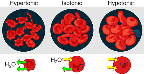
On their very own, cells can’t regulate the motion of water molecules throughout their membrane, so it is necessary that cells are uncovered to an setting during which the focus of solutes exterior of the cells (within the extracellular fluid) is the same as the focus of solutes contained in the cells (within the cytoplasm). Two options which have the identical focus of solutes are mentioned to be isotonic (equal rigidity). When cells and their extracellular environments are isotonic, the focus of water molecules is similar inside and outside the cells, and the cells preserve their regular form (and performance).
Osmosis happens when there’s an imbalance of solutes exterior of a cell versus contained in the cell. An answer that has the next focus of solutes than one other answer is claimed to be hypertonic, and water molecules are inclined to diffuse right into a hypertonic answer (Determine 3.1.6). Cells in a hypertonic answer will shrivel as water leaves the cell by way of osmosis. In distinction, an answer that has a decrease focus of solutes than one other answer is claimed to be hypotonic, and water molecules are inclined to diffuse out of a hypotonic answer. Cells in a hypotonic answer will tackle an excessive amount of water and swell, with the danger of ultimately bursting. A important facet of homeostasis in residing issues is to create an inner setting during which the entire physique’s cells are in an isotonic answer. Numerous organ programs, significantly the kidneys, work to take care of this homeostasis.
Energetic Transport
For the entire transport strategies described above, the cell expends no power. Membrane proteins that help within the passive transport of gear accomplish that with out the usage of ATP. Throughout main lively transport, ATP is required to maneuver a substance throughout a membrane, with the assistance of membrane protein, and in opposition to its focus gradient.
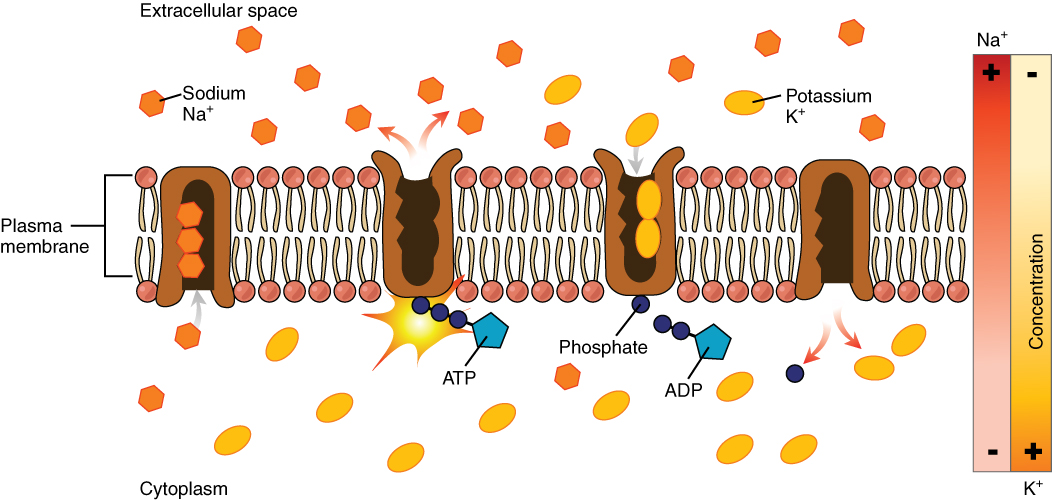
One of the widespread forms of lively transport entails proteins that function pumps. The phrase “pump” in all probability conjures up ideas of utilizing power to pump up the tire of a bicycle or a basketball. Equally, power from ATP is required for these membrane proteins to move substances—molecules or ions—throughout the membrane, in opposition to their focus gradients (from an space of low focus to an space of excessive focus).
The sodium-potassium pump, which can also be referred to as Na+/Okay+ ATPase, transports sodium out of a cell whereas transferring potassium into the cell. The Na+/Okay+ pump is a crucial ion pump discovered within the membranes of all cells. The exercise of those pumps in nerve cells is so nice that it accounts for almost all of their ATP utilization.
Energetic transport pumps may work along with different lively or passive transport programs to maneuver substances throughout the membrane. For instance, the sodium-potassium pump maintains a excessive focus of sodium ions exterior of the cell. Subsequently, if the cell wants sodium ions, all it has to do is open a passive sodium channel, because the focus gradient of the sodium ions will drive them to diffuse into the cell. On this means, the motion of an lively transport pump (the sodium-potassium pump) powers the passive transport of sodium ions by making a focus gradient. When lively transport powers the transport of one other substance on this means, it’s referred to as secondary lively transport.
Symporters are secondary lively transporters that transfer two substances in the identical path. For instance, the sodium-glucose symporter makes use of sodium ions to “pull” glucose molecules into the cell. Since cells retailer glucose for power, glucose is often at the next focus within the cell than exterior; nonetheless, as a result of motion of the sodium-potassium pump, sodium ions will simply diffuse into the cell when the symporter is opened. The flood of sodium ions by way of the symporter offers the power that permits glucose to maneuver by way of the symporter and into the cell, in opposition to its focus gradient.
Conversely, antiporters are secondary lively transport programs that transport substances in reverse instructions. For instance, the sodium-hydrogen ion antiporter makes use of the power from the inward flood of sodium ions to maneuver hydrogen ions (H+) out of the cell. The sodium-hydrogen antiporter is used to take care of the pH of the cell’s inside.
Different Types of Membrane Transport
Different types of lively transport don’t contain membrane carriers. Endocytosis (bringing “into the cell”) is the method of a cell ingesting materials by enveloping it in a portion of its cell membrane, after which pinching off that portion of membrane (Determine 3.1.8). As soon as pinched off, the portion of membrane and its contents turns into an unbiased, intracellular vesicle. A vesicle is a membranous sac—a spherical and hole organelle bounded by a lipid bilayer membrane. Endocytosis usually brings supplies into the cell that should to be damaged down or digested. Phagocytosis (“cell consuming”) is the endocytosis of huge particles. Many immune cells have interaction in phagocytosis of invading pathogens. Like little Pac-men, their job is to patrol physique tissues for undesirable matter, comparable to invading bacterial cells, phagocytize them, and digest them. In distinction to phagocytosis, pinocytosis (“cell consuming”) brings fluid containing dissolved substances right into a cell by way of membrane vesicles.
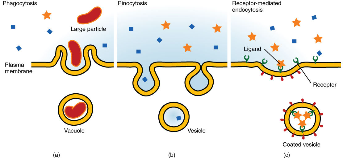
Phagocytosis and pinocytosis soak up giant parts of extracellular materials, and they’re sometimes not extremely selective within the substances they carry in. Cells regulate the endocytosis of particular substances by way of receptor-mediated endocytosis. Receptor-mediated endocytosis is endocytosis by a portion of the cell membrane which incorporates many receptors which might be particular for a sure substance. As soon as the floor receptors have sure ample quantities of the precise substance (the receptor’s ligand), the cell will endocytose the a part of the cell membrane containing the receptor-ligand complexes. Iron, a required element of hemoglobin, is endocytosed by pink blood cells on this means. Iron is sure to a protein referred to as transferrin within the blood. Particular transferrin receptors on pink blood cell surfaces bind the iron-transferrin molecules, and the cell endocytoses the receptor-ligand complexes.
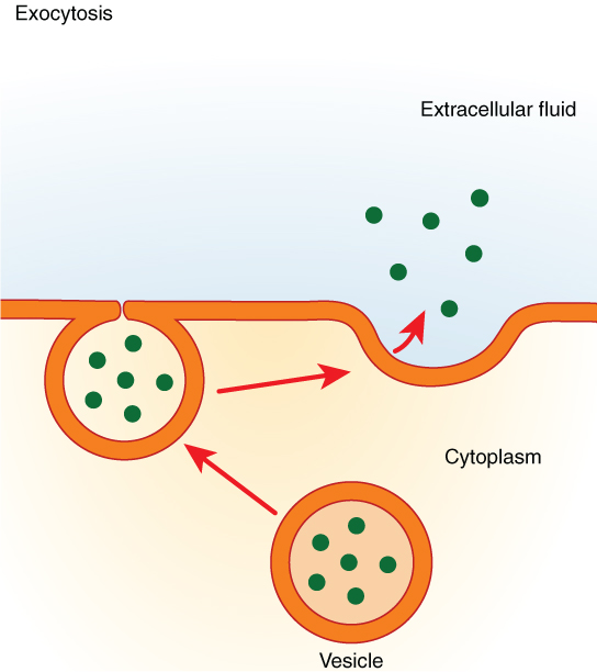
In distinction with endocytosis, exocytosis (taking “out of the cell”) is the method of a cell exporting materials utilizing vesicular transport (Determine 3.1.9). Many cells manufacture substances that should be secreted, like a manufacturing unit manufacturing a product for export. These substances are sometimes packaged into membrane-bound vesicles throughout the cell. When the vesicle membrane fuses with the cell membrane, the vesicle releases its contents into the interstitial fluid. The vesicle membrane then turns into a part of the cell membrane.
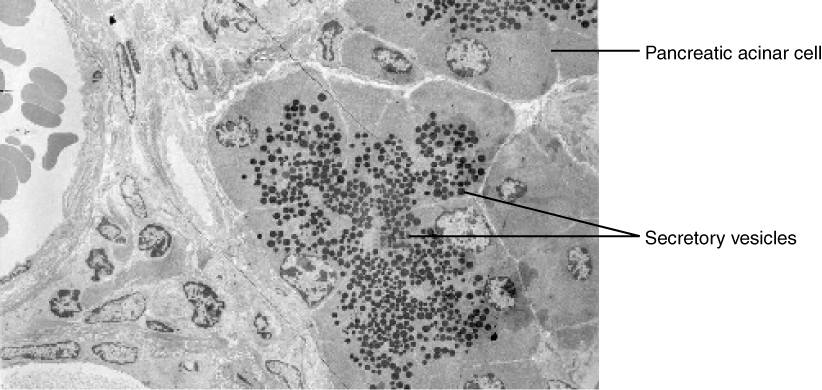
Particular examples of exocytosis embrace cells of the abdomen and pancreas producing and secreting digestive enzymes by way of exocytosis (Determine 3.1.10) and endocrine cells producing and secreting hormones which might be despatched all through the physique.
The addition of recent membrane to the plasma membrane is normally coupled with endocytosis in order that the cell will not be consistently enlarging. By these processes, the cell membrane is consistently renewing and altering as wanted by the cell.
Illnesses of the Cell: Cystic Fibrosis
Cystic fibrosis (CF) impacts roughly 30,000 individuals in the USA, with about 1,000 new instances reported every year. The genetic illness is most well-known for its injury to the lungs, inflicting respiration difficulties and power lung infections, but it surely additionally impacts the liver, pancreas, and intestines. Solely about 50 years in the past, the prognosis for kids born with CF was very grim—a life expectancy not often over 10 years. At this time, with advances in medical remedy, many CF sufferers stay into their 30s.
The signs of CF outcome from a malfunctioning membrane ion channel referred to as the Cystic Fibrosis Transmembrane Conductance Regulator, or CFTR. In wholesome individuals, the CFTR protein is an integral membrane protein that transports Cl– ions out of the cell. In an individual who has CF, the gene for the CFTR is mutated, thus, the cell manufactures a faulty channel protein that sometimes will not be included into the membrane, however is as a substitute degraded by the cell.
The CFTR requires ATP to be able to operate, making its Cl– transport a type of lively transport. This puzzled researchers for a very long time as a result of the Cl– ions are literally flowing down their focus gradient when transported out of cells. Energetic transport usually pumps ions in opposition to their focus gradient, however the CFTR presents an exception to this rule.
In regular lung tissue, the motion of Cl– out of the cell maintains a Cl–-rich, negatively charged setting instantly exterior of the cell. That is significantly vital within the epithelial lining of the respiratory system. Respiratory epithelial cells secrete mucus, which serves to entice mud, micro organism, and different particles. A cilium (plural = cilia) is likely one of the hair-like appendages discovered on sure cells. Cilia on the epithelial cells transfer the mucus and its trapped particles up the airways away from the lungs and towards the skin. With a purpose to be successfully moved upward, the mucus can’t be too viscous, somewhat, it should have a skinny, watery consistency. The transport of Cl– and the upkeep of an electronegative setting exterior of the cell attracts constructive ions comparable to Na+ to the extracellular area. The buildup of each Cl– and Na+ ions within the extracellular area creates solute-rich mucus, which has a low focus of water molecules. In consequence, by way of osmosis, water strikes from cells and extracellular matrix into the mucus, “thinning” it out. In a traditional respiratory system, that is how the mucus is saved sufficiently watered-down to be propelled out of the respiratory system.
If the CFTR channel is absent, Cl– ions aren’t transported out of the cell in sufficient numbers, thus stopping them from drawing constructive ions. The absence of ions within the secreted mucus leads to the shortage of a traditional water focus gradient. Thus, there is no such thing as a osmotic strain pulling water into the mucus. The ensuing mucus is thick and sticky, and the ciliated epithelia can’t successfully take away it from the respiratory system. Passageways within the lungs turn into blocked with mucus, together with the particles it carries. Bacterial infections happen extra simply as a result of bacterial cells aren’t successfully carried away from the lungs.
Chapter Assessment
The cell membrane offers a barrier across the cell, separating its inner parts from the extracellular setting. It’s composed of a phospholipid bilayer, with hydrophobic inner lipid “tails” and hydrophilic exterior phosphate “heads.” Numerous membrane proteins are scattered all through the bilayer, each inserted inside it and connected to it peripherally. The cell membrane is selectively permeable, permitting solely a restricted variety of supplies to diffuse by way of its lipid bilayer. All supplies that cross the membrane accomplish that utilizing passive (non-energy-requiring) or lively (energy-requiring) transport processes. Throughout passive transport, supplies transfer by easy diffusion or by facilitated diffusion by way of the membrane, down their focus gradient. Water passes by way of the membrane in a diffusion course of referred to as osmosis. Throughout lively transport, power is expended to help materials motion throughout the membrane in a path in opposition to their focus gradient. Energetic transport could happen with the assistance of protein pumps or by way of the usage of vesicles.
3.2 The Cytoplasm and Mobile Organelles
Studying Targets
By the top of this part, it is possible for you to to:
- Describe the construction and performance of the mobile organelles related to the endomembrane system, together with the endoplasmic reticulum, Golgi equipment, and lysosomes
- Describe the construction and performance of mitochondria and peroxisomes
- Clarify the three parts of the cytoskeleton, together with their composition and features
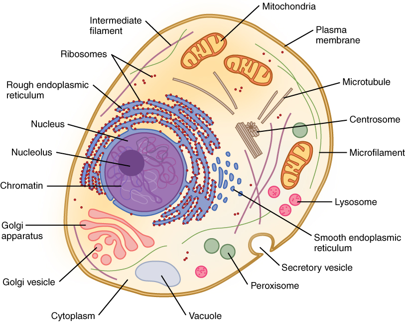
Now that you’ve realized that the cell membrane surrounds all cells, you’ll be able to dive within a prototypical human cell to find out about its inner parts and their features. All residing cells in multicellular organisms comprise an inner cytoplasmic compartment, and a nucleus throughout the cytoplasm. Cytosol, the jelly-like substance throughout the cell, offers the fluid medium needed for biochemical reactions. Eukaryotic cells, together with all animal cells, additionally comprise varied mobile organelles. An organelle (“little organ”) is considered one of a number of various kinds of membrane-enclosed our bodies within the cell, every performing a singular operate. Simply as the varied bodily organs work collectively in concord to carry out all of a human’s features, the various completely different mobile organelles work collectively to maintain the cell wholesome and performing all of its vital features. The organelles and cytosol, taken collectively, compose the cell’s cytoplasm. The nucleus is a cell’s central organelle, which incorporates the cell’s DNA (Determine 3.2.1).
Organelles of the Endomembrane System
A set of three main organelles collectively type a system throughout the cell referred to as the endomembrane system. These organelles work collectively to carry out varied mobile jobs, together with the duty of manufacturing, packaging, and exporting sure mobile merchandise. The organelles of the endomembrane system embrace the endoplasmic reticulum, Golgi equipment, and vesicles.
Endoplasmic Reticulum
The endoplasmic reticulum (ER) is a system of channels that’s steady with the nuclear membrane (or “envelope”) protecting the nucleus and composed of the identical lipid bilayer materials. The ER could be considered a collection of winding thoroughfares much like the waterway canals in Venice. The ER offers passages all through a lot of the cell that operate in transporting, synthesizing, and storing supplies. The winding construction of the ER leads to a big membranous floor space that helps its many features (Determine 3.2.2).
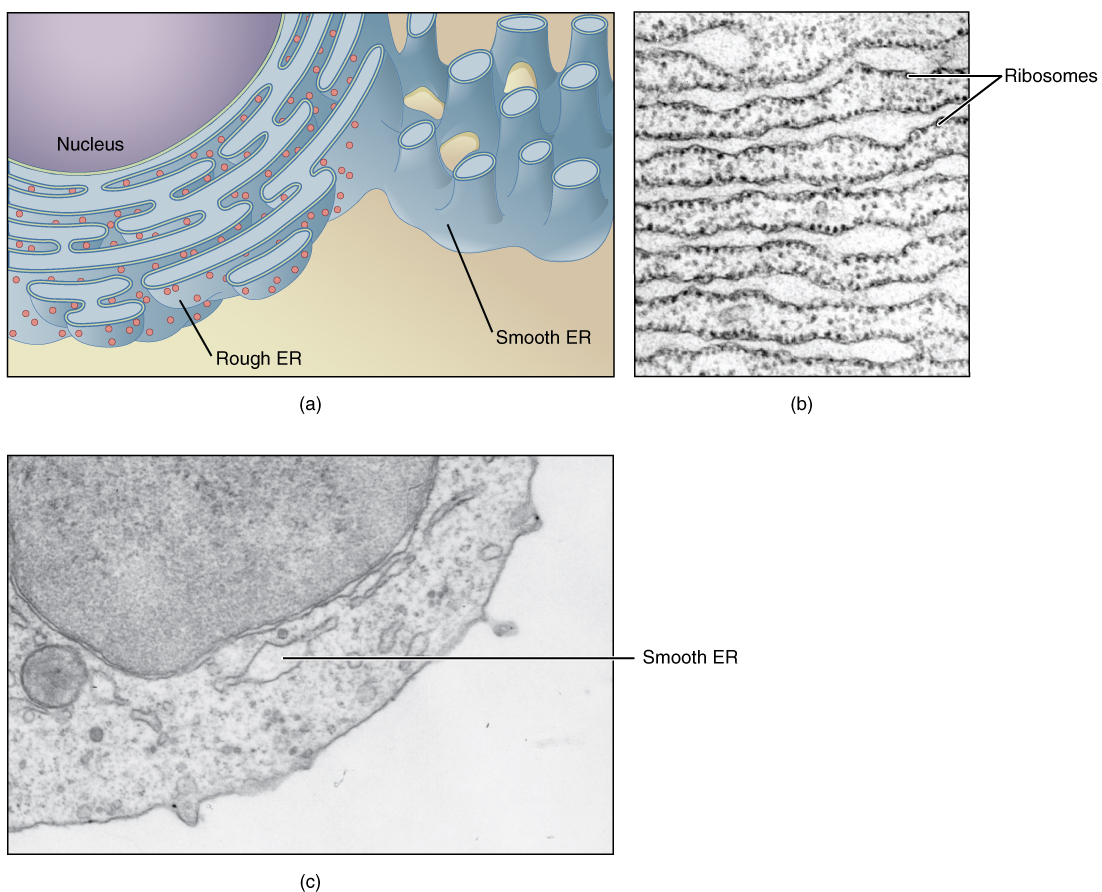
Endoplasmic reticulum can exist in two kinds: tough ER and clean ER. These two forms of ER carry out some very completely different features and could be present in very completely different quantities relying on the kind of cell. Tough ER (RER) is so-called as a result of its membrane is dotted with embedded granules—organelles referred to as ribosomes, giving the RER a bumpy look. A ribosome is an organelle that serves as the location of protein synthesis. It’s composed of two ribosomal RNA subunits that wrap round mRNA to start out the method of translation, adopted by protein synthesis. Easy ER (SER) lacks these ribosomes.
One of many primary features of the sleek ER is within the synthesis of lipids. The sleek ER synthesizes phospholipids, the principle element of organic membranes, in addition to steroid hormones. For that reason, cells that produce giant portions of such hormones, comparable to these of the feminine ovaries and male testes, comprise giant quantities of clean ER. Along with lipid synthesis, the sleek ER additionally sequesters (i.e., shops) and regulates the focus of mobile Ca++, a operate extraordinarily vital in cells of the nervous system the place Ca++ is the set off for neurotransmitter launch. The sleek ER moreover metabolizes some carbohydrates and performs a cleansing function, breaking down sure toxins.
In distinction with the sleek ER, the first job of the tough ER is the synthesis and modification of proteins destined for the cell membrane or for export from the cell. For this protein synthesis, many ribosomes connect to the ER (giving it the studded look of tough ER). Sometimes, a protein is synthesized throughout the ribosome and launched contained in the channel of the tough ER, the place sugars could be added to it (by a course of referred to as glycosylation) earlier than it’s transported inside a vesicle to the subsequent stage within the packaging and delivery course of: the Golgi equipment.
The Golgi Equipment
The Golgi equipment is accountable for sorting, modifying, and delivery off the merchandise that come from the tough ER, very similar to a post-office. The Golgi equipment appears like stacked flattened discs, nearly like stacks of oddly formed pancakes. Just like the ER, these discs are membranous. The Golgi equipment has two distinct sides, every with a unique function. One aspect of the equipment receives merchandise in vesicles. These merchandise are sorted by way of the equipment after which they’re launched from the other aspect after being repackaged into new vesicles. If the product is to be exported from the cell, the vesicle migrates to the cell floor and fuses to the cell membrane, and the cargo is secreted (Determine 3.2.3).
Lysosomes
Among the protein merchandise packaged by the Golgi embrace digestive enzymes that should stay contained in the cell to be used in breaking down sure supplies. The enzyme-containing vesicles launched by the Golgi could type new lysosomes, or fuse with present, lysosomes. A lysosome is an organelle that incorporates enzymes that break down and digest unneeded mobile parts, comparable to a broken organelle. (A lysosome is much like a wrecking crew that takes down previous and unsound buildings in a neighborhood.) Autophagy (“self-eating”) is the method of a cell digesting its personal buildings. Lysosomes are additionally vital for breaking down overseas materials. For instance, when sure immune protection cells (white blood cells) phagocytize micro organism, the bacterial cell is transported right into a lysosome and digested by the enzymes inside. As one may think, such phagocytic protection cells comprise giant numbers of lysosomes.
Below sure circumstances, lysosomes carry out a extra grand and dire operate. Within the case of broken or unhealthy cells, lysosomes could be triggered to open up and launch their digestive enzymes into the cytoplasm of the cell, killing the cell. This “self-destruct” mechanism known as autolysis, and makes the method of cell dying managed (a mechanism referred to as “apoptosis”).
Organelles for Power Manufacturing and Detoxing
Along with the roles carried out by the endomembrane system, the cell has many different vital features. Simply as you will need to eat vitamins to supply your self with power, so should every of your cells soak up vitamins, a few of which convert to chemical power that can be utilized to energy biochemical reactions. One other vital operate of the cell is cleansing. People soak up all kinds of poisons from the setting and likewise produce dangerous chemical compounds as byproducts of mobile processes. Cells referred to as hepatocytes within the liver detoxify many of those toxins.
Mitochondria
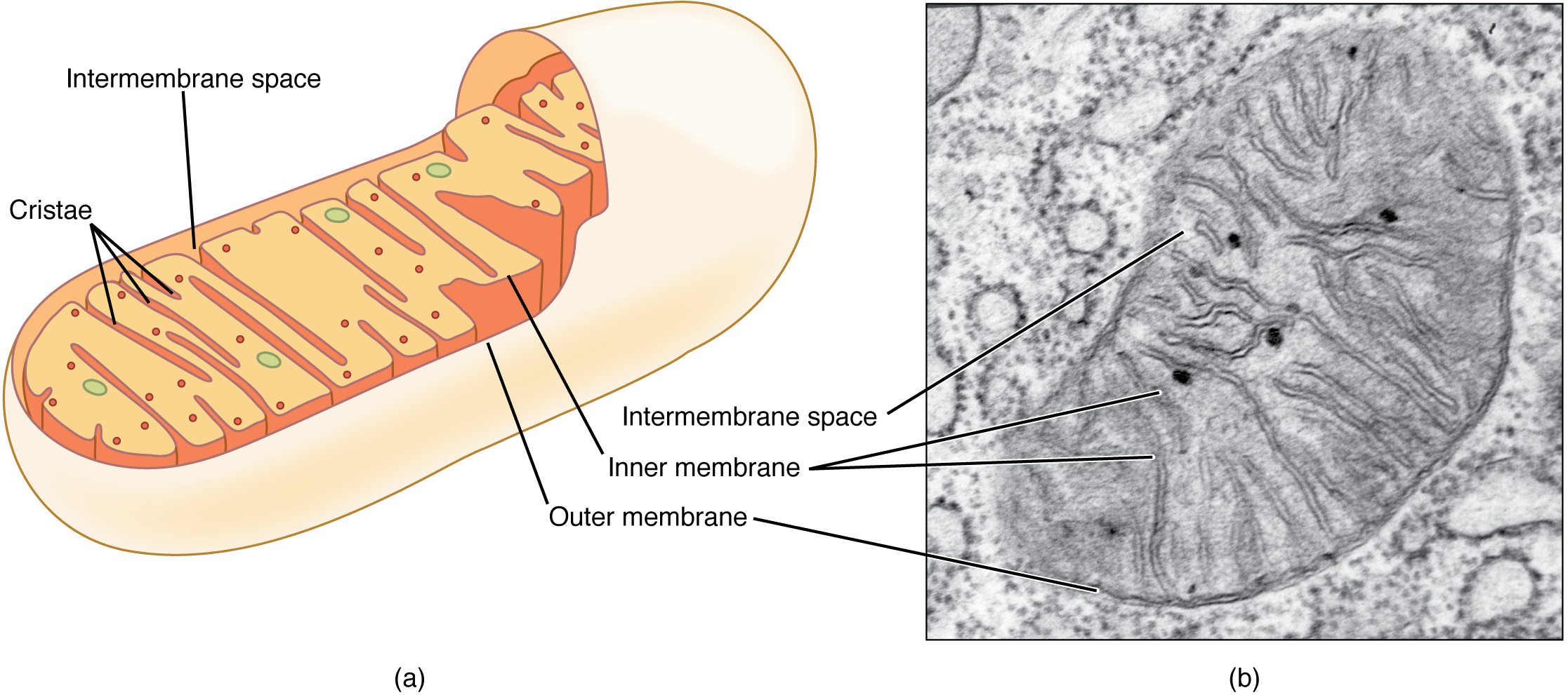
A mitochondrion (plural = mitochondria) is a membranous, bean-shaped organelle that’s the “power transformer” of the cell. Mitochondria encompass an outer lipid bilayer membrane in addition to an extra inside lipid bilayer membrane (Determine 3.2.4). The inside membrane is extremely folded into winding buildings with quite a lot of floor space, referred to as cristae. It’s alongside this inside membrane {that a} collection of proteins, enzymes, and different molecules carry out the biochemical reactions of mobile respiration. These reactions convert power saved in nutrient molecules (comparable to glucose) into adenosine triphosphate (ATP), which offers usable mobile power to the cell. Cells use ATP consistently, and so the mitochondria are consistently at work. Oxygen molecules are required throughout mobile respiration, which is why you will need to consistently breathe it in. One of many organ programs within the physique that makes use of big quantities of ATP is the muscular system as a result of ATP is required to maintain muscle contraction. In consequence, muscle cells are packed filled with mitochondria. Nerve cells additionally want giant portions of ATP to run their sodium-potassium pumps. Subsequently, a person neuron can be loaded with over a thousand mitochondria. Alternatively, a bone cell, which isn’t practically as metabolically-active, may solely have a pair hundred mitochondria.
Peroxisomes
Like lysosomes, a peroxisome is a membrane-bound mobile organelle that incorporates principally enzymes (Determine 3.2.5). Peroxisomes carry out a few completely different features, together with lipid metabolism and chemical cleansing. In distinction to the digestive enzymes present in lysosomes, the enzymes inside peroxisomes serve to switch hydrogen atoms from varied molecules to oxygen, producing hydrogen peroxide (H2O2). On this means, peroxisomes neutralize poisons comparable to alcohol. With a purpose to recognize the significance of peroxisomes, it’s needed to grasp the idea of reactive oxygen species.
Reactive oxygen species (ROS) comparable to peroxides and free radicals are the extremely reactive merchandise of many regular mobile processes, together with the mitochondrial reactions that produce ATP and oxygen metabolism. Examples of ROS embrace the hydroxyl radical OH, H2O2, and superoxide (O2−). Some ROS are vital for sure mobile features, comparable to cell signaling processes and immune responses in opposition to overseas substances. Free radicals are reactive as a result of they comprise free unpaired electrons; they will simply oxidize different molecules all through the cell, inflicting mobile injury and even cell dying. Free radicals are thought to play a task in lots of damaging processes within the physique, from most cancers to coronary artery illness.
Peroxisomes, however, oversee reactions that neutralize free radicals. Peroxisomes produce giant quantities of the poisonous H2O2 within the course of, but in addition comprise enzymes that convert H2O2 into water and oxygen. These byproducts are safely launched into the cytoplasm. Like miniature sewage remedy vegetation, peroxisomes neutralize dangerous toxins in order that they don’t wreak havoc within the cells. The liver is the organ primarily accountable for detoxifying the blood earlier than it travels all through the physique, and liver cells comprise an exceptionally excessive variety of peroxisomes.
Protection mechanisms comparable to cleansing throughout the peroxisome and sure mobile antioxidants serve to neutralize many of those molecules. Some nutritional vitamins and different substances, discovered primarily in vegatables and fruits, have antioxidant properties. Antioxidants work by being oxidized themselves, halting the damaging response cascades initiated by the free radicals. Typically although, ROS accumulate past the capability of such defenses.
Oxidative stress is the time period used to explain injury to mobile parts attributable to ROS. Attributable to their distinctive unpaired electrons, ROS can set off chain reactions the place they take away electrons from different molecules, which then turn into oxidized and reactive; they do the identical to different molecules, inflicting a sequence response. ROS could cause everlasting injury to mobile lipids, proteins, carbohydrates, and nucleic acids. Broken DNA can result in genetic mutations and even most cancers. A mutation is a change within the nucleotide sequence in a gene inside a cell’s DNA, probably altering the protein coded by that gene. Different illnesses believed to be triggered or exacerbated by ROS embrace Alzheimer’s illness, cardiovascular illnesses, diabetes, Parkinson’s illness, arthritis, Huntington’s illness, and schizophrenia, amongst many others. It’s noteworthy that these illnesses are largely age-related. Many scientists imagine that oxidative stress is a significant contributor to the getting older course of.
Ageing and the…Cell: The Free Radical IdeaThe free radical principle on getting older was initially proposed within the Nineteen Fifties, and nonetheless stays underneath debate. Typically talking, the free radical principle of getting older means that gathered mobile injury from oxidative stress contributes to the physiological and anatomical results of getting older. There are two considerably completely different variations of this principle: one states that the getting older course of itself is a results of oxidative injury, and the opposite states that oxidative injury causes age-related illnesses and problems. The latter model of the idea is extra extensively accepted than the previous. Nonetheless, many strains of proof counsel that oxidative injury does contribute to the getting older course of. Analysis has proven that lowering oxidative injury can lead to an extended lifespan in sure organisms comparable to yeast, worms, and fruit flies. Conversely, growing oxidative injury can shorten the lifespan of mice and worms. Apparently, a manipulation referred to as calorie-restriction (reasonably proscribing the caloric consumption) has been proven to extend life span in some laboratory animals. It’s believed that this enhance is not less than partly resulting from a discount of oxidative stress. Nonetheless, a long-term research of primates with calorie-restriction confirmed no enhance of their lifespan. An excessive amount of extra analysis can be required to raised perceive the hyperlink between reactive oxygen species and getting older.
The Cytoskeleton
Very like the bony skeleton structurally helps the human physique, the cytoskeleton helps the cells to take care of their structural integrity. The cytoskeleton is a bunch of fibrous proteins that present structural assist for cells, however that is solely one of many features of the cytoskeleton. Cytoskeletal parts are additionally important for cell motility, cell replica, and transportation of gear throughout the cell.
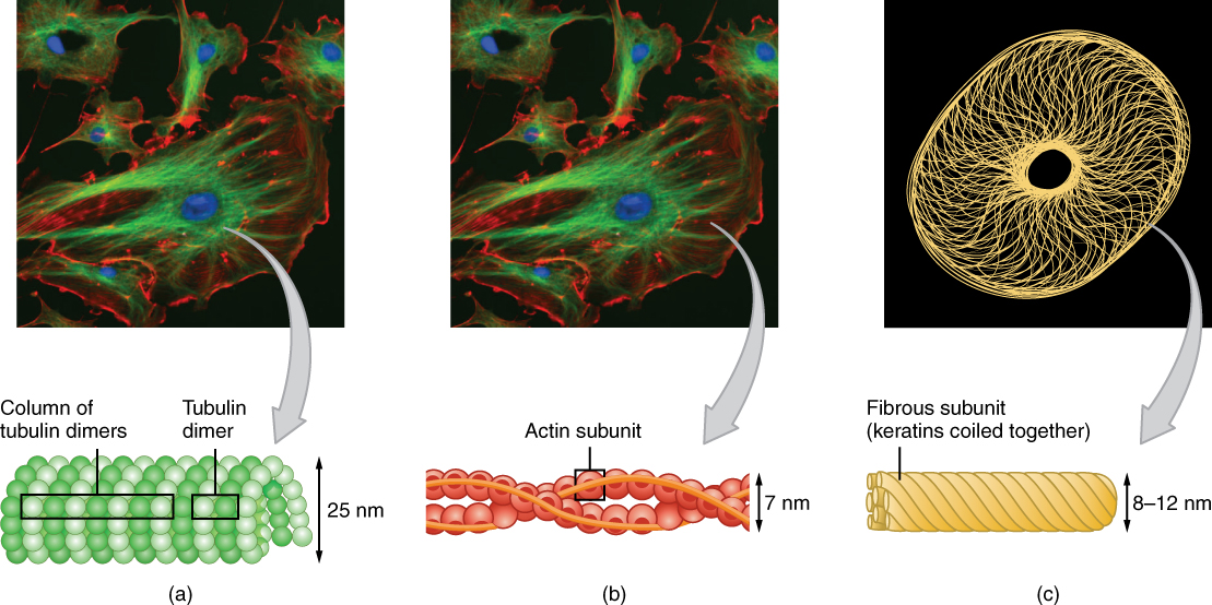
The cytoskeleton kinds a fancy thread-like community all through the cell consisting of three completely different sorts of protein-based filaments: microfilaments, intermediate filaments, and microtubules (Determine 3.2.6). The thickest of the three is the microtubule, a structural filament composed of subunits of a protein referred to as tubulin. Microtubules preserve cell form and construction, assist resist compression of the cell, and play a task in positioning the organelles throughout the cell. Microtubules additionally make up two forms of mobile appendages vital for movement: cilia and flagella. Cilia are discovered on many cells of the physique, together with the epithelial cells that line the airways of the respiratory system. Cilia transfer rhythmically; they beat consistently, transferring waste supplies comparable to mud, mucus, and micro organism upward by way of the airways, away from the lungs and towards the mouth. Beating cilia on cells within the feminine fallopian tubes transfer egg cells from the ovary in direction of the uterus. A flagellum (plural = flagella) is an appendage bigger than a cilium and specialised for cell locomotion. The one flagellated cell in people is the sperm cell that should propel itself in direction of feminine egg cells.
An important operate of microtubules is to set the paths (considerably like railroad tracks) alongside the place the genetic materials could be pulled (a course of requiring ATP) throughout cell division, so that every new daughter cell receives the suitable set of chromosomes. Two quick, equivalent microtubule buildings referred to as centrioles are discovered close to the nucleus of cells. A centriole can function the mobile origin level for microtubules extending outward as cilia or flagella or can help with the separation of DNA throughout cell division. Microtubules develop out from the centrioles by including extra tubulin subunits, like including extra hyperlinks to a sequence.
In distinction with microtubules, the microfilament is a thinner kind of cytoskeletal filament (see Determine 3.2.6b). Actin, a protein that kinds chains, is the first element of those microfilaments. Actin fibers, twisted chains of actin filaments, represent a big element of muscle tissue and, together with the protein myosin, are accountable for muscle contraction. Like microtubules, actin filaments are lengthy chains of single subunits (referred to as actin subunits). In muscle cells, these lengthy actin strands, referred to as skinny filaments, are “pulled” by thick filaments of the myosin protein to contract the cell.
Actin additionally has an vital function throughout cell division. When a cell is about to separate in half throughout cell division, actin filaments work with myosin to create a cleavage furrow that ultimately splits the cell down the center, forming two new cells from the unique cell.
The ultimate cytoskeletal filament is the intermediate filament. As its title would counsel, an intermediate filament is a filament intermediate in thickness between the microtubules and microfilaments (see Determine 3.2.6c). Intermediate filaments are made up of lengthy fibrous subunits of a protein referred to as keratin which might be wound collectively just like the threads that compose a rope. Intermediate filaments, in live performance with the microtubules, are vital for sustaining cell form and construction. Not like the microtubules, which resist compression, intermediate filaments resist rigidity—the forces that pull aside cells. There are lots of instances during which cells are susceptible to rigidity, comparable to when epithelial cells of the pores and skin are compressed, tugging them in several instructions. Intermediate filaments assist anchor organelles collectively inside a cell and likewise hyperlink cells to different cells by forming particular cell-to-cell junctions.
Chapter Assessment
The inner setting of a residing cell is made up of a fluid, jelly-like substance referred to as cytosol, which consists primarily of water, but in addition incorporates varied dissolved vitamins and different molecules. The cell incorporates an array of mobile organelles, every one performing a singular operate and serving to to take care of the well being and exercise of the cell. The cytosol and organelles collectively compose the cell’s cytoplasm. Most organelles are surrounded by a lipid membrane much like the cell membrane of the cell. The endoplasmic reticulum (ER), Golgi equipment, and lysosomes share a useful connectivity and are collectively known as the endomembrane system. There are two forms of ER: clean and tough. Whereas the sleek ER performs many features, together with lipid synthesis and ion storage, the tough ER is principally accountable for protein synthesis utilizing its related ribosomes. The tough ER sends newly made proteins to the Golgi equipment the place they’re modified and packaged for supply to numerous areas inside or exterior of the cell. A few of these protein merchandise are enzymes destined to interrupt down undesirable materials and are packaged as lysosomes to be used contained in the cell.
Cells additionally comprise mitochondria and peroxisomes, that are the organelles accountable for producing the cell’s power provide and detoxifying sure chemical compounds, respectively. Biochemical reactions inside mitochondria remodel energy-carrying molecules into the usable type of mobile power generally known as ATP. Peroxisomes comprise enzymes that remodel dangerous substances comparable to free radicals into oxygen and water. Cells additionally comprise a miniaturized “skeleton” of protein filaments that stretch all through its inside. Three completely different sorts of filaments compose this cytoskeleton (so as of accelerating thickness): microfilaments, intermediate filaments, and microtubules. Every cytoskeletal element performs distinctive features in addition to offers a supportive framework for the cell.
3.3 The Nucleus and DNA Replication
Studying Targets
By the top of this part, it is possible for you to to:
- Describe the construction and options of the nuclear membrane
- Listing the contents of the nucleus
- Clarify the group of the DNA molecule throughout the nucleus
- Describe the method of DNA replication
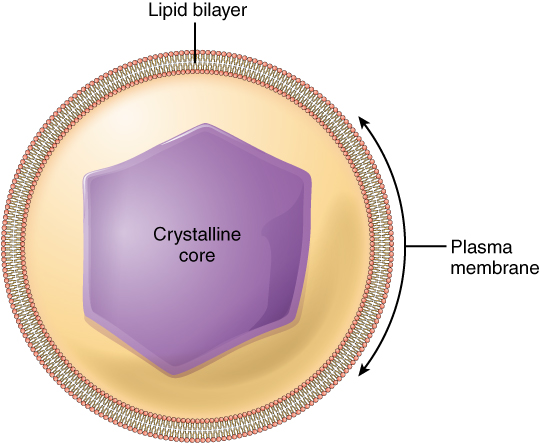
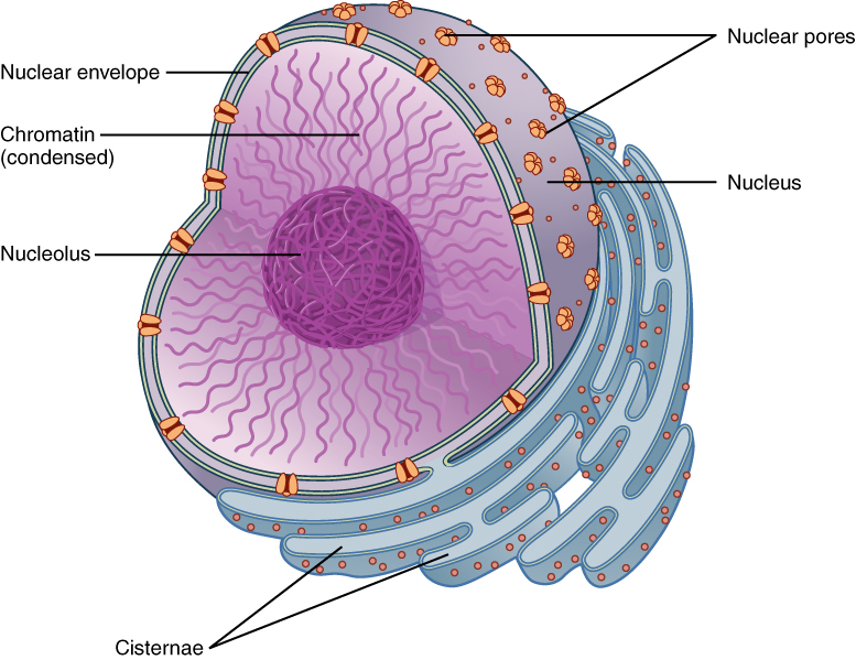
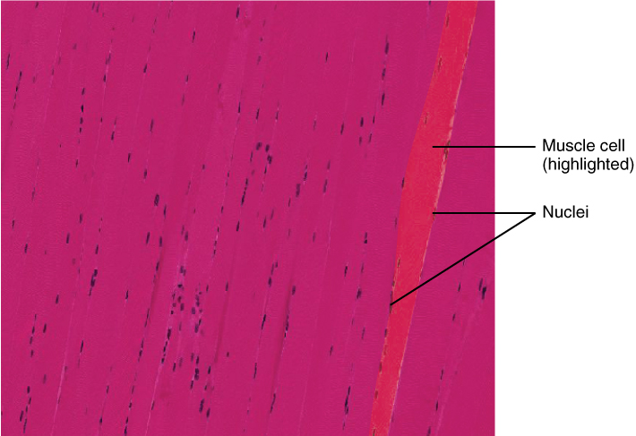
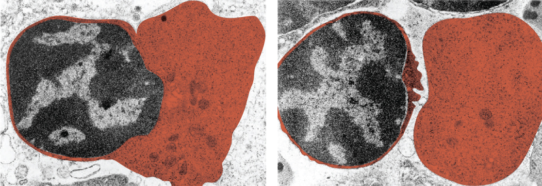
The nucleus is the most important and most outstanding of a cell’s organelles (Determine 3.3.1). The nucleus is usually thought of the management heart of the cell as a result of it shops the entire genetic directions for manufacturing proteins. Apparently, some cells within the physique, comparable to muscle cells, comprise multiple nucleus (Determine 3.3.2), which is named multinucleated. Different cells, comparable to mammalian pink blood cells (RBCs), don’t comprise nuclei in any respect. RBCs eject their nuclei as they mature, making area for the big numbers of hemoglobin molecules that carry oxygen all through the physique (Determine 3.3.3). With out nuclei, the life span of RBCs is brief, and so the physique should produce new ones consistently.
Contained in the nucleus lies the blueprint that dictates every part a cell will do and the entire merchandise it can make. This info is saved inside DNA. The nucleus sends “instructions” to the cell by way of molecular messengers that translate the knowledge from the DNA. Every cell in your physique (aside from germ cells) incorporates the entire set of your DNA. When a cell divides, the DNA should be duplicated so that every new cell receives a full complement of DNA. The next part will discover the construction of the nucleus and its contents, in addition to the method of DNA replication.
Group of the Nucleus and its DNA
Like most different mobile organelles, the nucleus is surrounded by a membrane referred to as the nuclear envelope. This membranous protecting consists of two adjoining lipid bilayers with a skinny fluid area in between them. Spanning these two bilayers are nuclear pores. A nuclear pore is a tiny passageway for the passage of proteins, RNA, and solutes between the nucleus and the cytoplasm. Proteins referred to as pore complexes lining the nuclear pores regulate the passage of supplies into and out of the nucleus.
Contained in the nuclear envelope is a gel-like nucleoplasm with solutes that embrace the constructing blocks of nucleic acids. There additionally is usually a dark-staining mass usually seen underneath a easy gentle microscope, referred to as a nucleolus (plural = nucleoli). The nucleolus is a area of the nucleus that’s accountable for manufacturing the RNA needed for building of ribosomes. As soon as synthesized, newly made ribosomal subunits exit the cell’s nucleus by way of the nuclear pores.
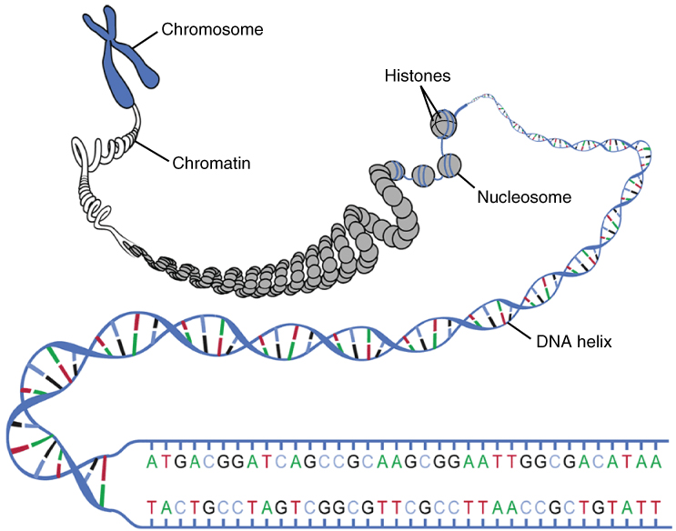
The genetic directions which might be used to construct and preserve an organism are organized in an orderly method in strands of DNA. Throughout the nucleus are threads of chromatin composed of DNA and related proteins (Determine 3.3.4). Alongside the chromatin threads, the DNA is wrapped round a set of histone proteins. A nucleosome is a single, wrapped DNA-histone complicated. A number of nucleosomes alongside the whole molecule of DNA appear as if a beaded necklace, during which the string is the DNA and the beads are the related histones. When a cell is within the technique of division, the chromatin condenses into chromosomes, in order that the DNA could be safely transported to the “daughter cells.” The chromosome consists of DNA and proteins; it’s the condensed type of chromatin. It’s estimated that people have nearly 22,000 genes distributed on 46 chromosomes.
DNA Replication
To ensure that an organism to develop, develop, and preserve its well being, cells should reproduce themselves by dividing to supply two new daughter cells, every with the total complement of DNA as discovered within the authentic cell. Billions of recent cells are produced in an grownup human on daily basis. Solely only a few cell sorts within the physique don’t divide, together with nerve cells, skeletal muscle fibers, and cardiac muscle cells. The division time of various cell sorts varies. Epithelial cells of the pores and skin and gastrointestinal lining, for example, divide very continuously to interchange these which might be consistently being rubbed off of the floor by friction.
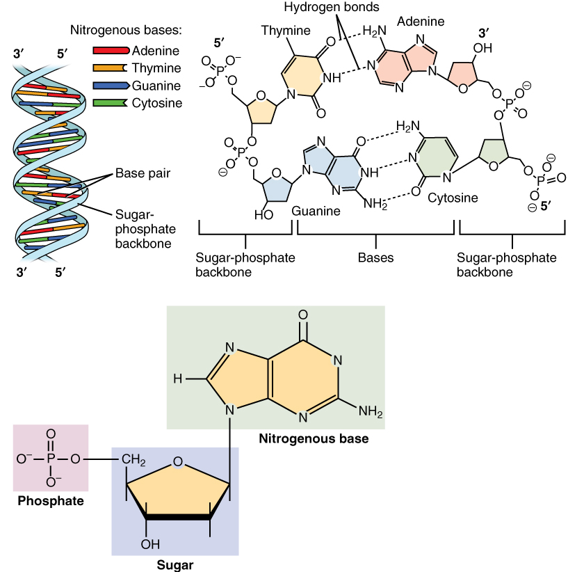
A DNA molecule is product of two strands that “complement” one another within the sense that the molecules that compose the strands match collectively and bind to one another, making a double-stranded molecule that appears very similar to an extended, twisted ladder. Both sides rail of the DNA ladder consists of alternating sugar and phosphate teams (Determine 3.3.5). The 2 sides of the ladder aren’t equivalent, however are complementary. These two backbones are bonded to one another throughout pairs of protruding bases, every bonded pair forming one “rung,” or cross member. The 4 DNA bases are adenine (A), thymine (T), cytosine (C), and guanine (G). Due to their form and cost, the 2 bases that compose a pair all the time bond collectively. Adenine all the time binds with thymine, and cytosine all the time binds with guanine. The actual sequence of bases alongside the DNA molecule determines the genetic code. Subsequently, if the 2 complementary strands of DNA had been pulled aside, you would infer the order of the bases in a single strand from the bases within the different, complementary strand. For instance, if one strand has a area with the sequence AGTGCCT, then the sequence of the complementary strand could be TCACGGA.
DNA replication is the copying of DNA that happens earlier than cell division can happen. After quite a lot of debate and experimentation, the overall technique of DNA replication was deduced in 1958 by two scientists in California, Matthew Meselson and Franklin Stahl. This technique is illustrated in Determine 3.3.6 and described under.
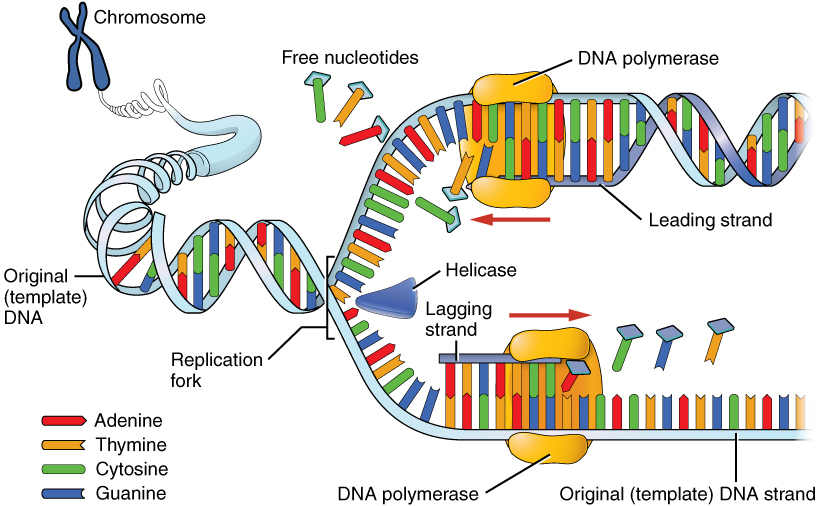
Stage 1: Initiation. The 2 complementary strands are separated, very similar to unzipping a zipper. Particular enzymes, together with helicase, untwist and separate the 2 strands of DNA.
Stage 2: Elongation. Every strand turns into a template alongside which a brand new complementary strand is constructed. DNA polymerase brings within the right bases to enhance the template strand, synthesizing a brand new strand base by base. A DNA polymerase is an enzyme that provides free nucleotides to the top of a sequence of DNA, making a brand new double strand. This rising strand continues to be constructed till it has totally complemented the template strand.
Stage 3: Termination. As soon as the 2 authentic strands are sure to their very own, completed, complementary strands, DNA replication is stopped and the 2 new equivalent DNA molecules are full.
Every new DNA molecule incorporates one strand from the unique molecule and one newly synthesized strand. The time period for this mode of replication is “semiconservative,” as a result of half of the unique DNA molecule is conserved in every new DNA molecule. This course of continues till the cell’s total genome, the whole complement of an organism’s DNA, is replicated. As you may think, it is extremely vital that DNA replication happen exactly in order that new cells within the physique comprise the very same genetic materials as their father or mother cells. Errors made throughout DNA replication, such because the unintended addition of an inappropriate nucleotide, have the potential to render a gene dysfunctional or ineffective. Luckily, there are mechanisms in place to reduce such errors. A DNA proofreading course of enlists the assistance of particular enzymes that scan the newly synthesized molecule for errors and corrects them. As soon as the method of DNA replication is full, the cell is able to divide. You’ll discover the method of cell division later within the chapter.
Chapter Assessment
The nucleus is the command heart of the cell, containing the genetic directions for the entire supplies a cell will make (and thus all of its features it might carry out). The nucleus is encased inside a membrane of two interconnected lipid bilayers, side-by-side. This nuclear envelope is studded with protein-lined pores that permit supplies to be trafficked into and out of the nucleus. The nucleus incorporates a number of nucleoli, which function websites for ribosome synthesis. The nucleus homes the genetic materials of the cell: DNA. DNA is often discovered as a loosely contained construction referred to as chromatin throughout the nucleus, the place it’s wound up and related to a wide range of histone proteins. When a cell is about to divide, the chromatin coils tightly and condenses to type chromosomes.
There’s a pool of cells consistently dividing inside your physique. The result’s billions of recent cells being created every day. Earlier than any cell is able to divide, it should replicate its DNA so that every new daughter cell will obtain a precise copy of the organism’s genome. A wide range of enzymes are enlisted throughout DNA replication. These enzymes unwind the DNA molecule, separate the 2 strands, and help with the constructing of complementary strands alongside every father or mother strand. The unique DNA strands function templates from which the nucleotide sequence of the brand new strands are decided and synthesized. When replication is accomplished, two equivalent DNA molecules exist. Each incorporates one authentic strand and one newly synthesized complementary strand.
3.4 Protein Synthesis
Studying Targets
Primary Goal
- Clarify the method by which a cell builds proteins utilizing the DNA code
By the top of this part, it is possible for you to to:
- Clarify how the genetic code inside DNA determines the proteins shaped
- Describe the method of transcription
- Clarify the method of translation
- Focus on the operate of ribosomes
It was talked about earlier that DNA offers a “blueprint” for the cell construction and physiology. This refers to the truth that DNA incorporates the knowledge needed for the cell to construct one crucial kind of molecule: the protein. Most structural parts of the cell are made up, not less than partly, by proteins and just about all of the features {that a} cell carries out are accomplished with the assistance of proteins. One of the vital lessons of proteins is enzymes, which assist pace up needed biochemical reactions that happen contained in the cell. A few of these important biochemical reactions embrace constructing bigger molecules from smaller parts (comparable to what happens throughout DNA replication or synthesis of microtubules) and breaking down bigger molecules into smaller parts (comparable to when harvesting chemical power from nutrient molecules). Regardless of the mobile course of could also be, it’s nearly certain to contain proteins. Simply because the cell’s genome describes its full complement of DNA, a cell’s proteome is its full complement of proteins. Protein synthesis begins with genes. A gene is a useful phase of DNA that gives the genetic info needed to construct a protein. Every explicit gene offers the code essential to assemble a selected protein. Gene expression, which transforms the knowledge coded in a gene to a remaining gene product, in the end dictates the construction and performance of a cell by figuring out which proteins are made.
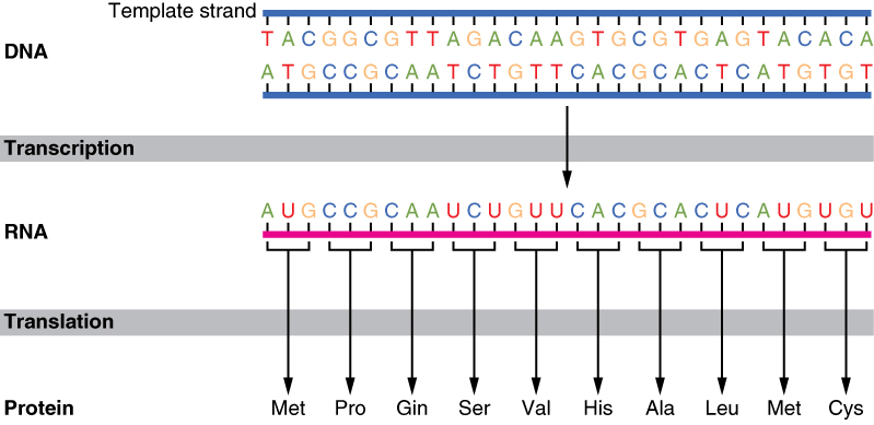
The interpretation of genes works within the following means. Recall that proteins are polymers, or chains, of many amino acid constructing blocks. The sequence of bases in a gene (that’s, its sequence of A, T, C, G nucleotides) interprets to an amino acid sequence. A triplet is a bit of three DNA bases in a row that codes for a particular amino acid. For instance, the DNA triplet CAC (cytosine, adenine, and cytosine) specifies the amino acid valine. Subsequently, a gene, which consists of a number of triplets in a singular sequence, offers the code to construct a complete protein, with a number of amino acids within the correct sequence (Determine 3.4.1). The mechanism by which cells flip the DNA code right into a protein product is a two-step course of, with an RNA molecule because the intermediate.
From DNA to RNA: Transcription
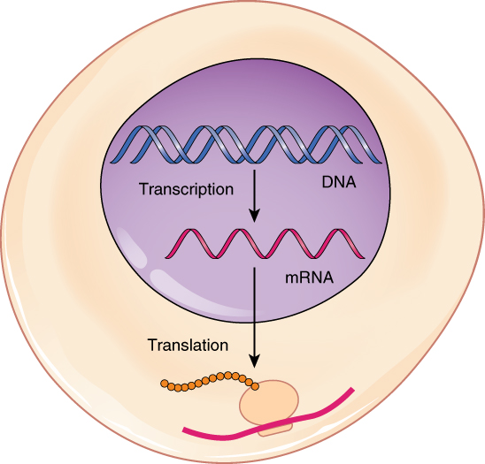
DNA is housed throughout the nucleus, and protein synthesis takes place within the cytoplasm, thus there should be some type of intermediate messenger that leaves the nucleus and manages protein synthesis. This intermediate messenger is messenger RNA (mRNA), (Determine 3.29), a single-stranded nucleic acid that carries a duplicate of the genetic code for a single gene out of the nucleus and into the cytoplasm the place it’s used to supply proteins.
There are a number of various kinds of RNA, every having completely different features within the cell. The construction of RNA is much like DNA with a couple of small exceptions. For one factor, not like DNA, most forms of RNA, together with mRNA, are single-stranded and comprise no complementary strand. Second, the ribose sugar in RNA incorporates an extra oxygen atom in contrast with DNA. Lastly, as a substitute of the bottom thymine, RNA incorporates the bottom uracil. Which means adenine will all the time pair up with uracil in the course of the protein synthesis course of.
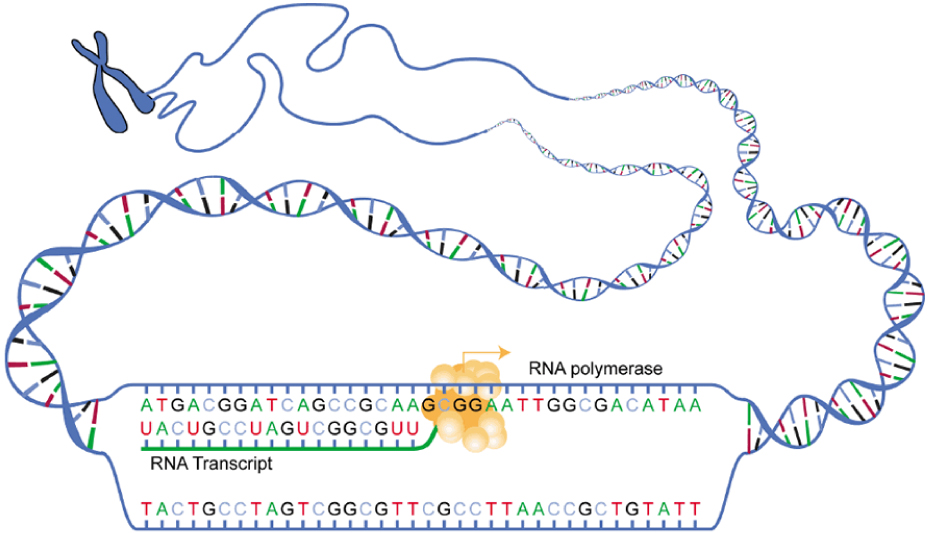
Gene expression begins with the method referred to as transcription, which is the synthesis of a strand of mRNA that’s complementary to the gene of curiosity. This course of known as transcription as a result of the mRNA is sort of a transcript, or copy, of the gene’s DNA code. Transcription begins in a vogue considerably like DNA replication, in {that a} area of DNA unwinds and the 2 strands separate, nonetheless, solely that small portion of the DNA can be cut up aside. The triplets throughout the gene on this part of the DNA molecule are used because the template to transcribe the complementary strand of RNA (Figure 3.4.2). A codon is a three-base sequence of mRNA, so-called as a result of they instantly encode amino acids. Like DNA replication, there are three levels to transcription: initiation, elongation, and termination.
Within the first of the 2 levels of constructing protein from DNA, a gene on the DNA molecule is transcribed right into a complementary mRNA molecule.
Stage 1: Initiation. A area initially of the gene referred to as a promoter—a selected sequence of nucleotides—triggers the beginning of transcription.
Stage 2: Elongation. Transcription begins when RNA polymerase unwinds the DNA phase. One strand, known as the coding strand, turns into the template with the genes to be coded. The polymerase then aligns the proper nucleic acid (A, C, G, or U) with its complementary base on the coding strand of DNA. RNA polymerase is an enzyme that provides new nucleotides to a rising strand of RNA. This course of builds a strand of mRNA.
Stage 3: Termination. When the polymerase has reached the top of the gene, considered one of three particular triplets (UAA, UAG, or UGA) codes a “cease” sign, which triggers the enzymes to terminate transcription and launch the mRNA transcript.
The transcription course of is regulated by a category of proteins referred to as transcription components, which bind to the gene sequence and both promote or inhibit their transcription. (transfer Determine 3.35 right here).
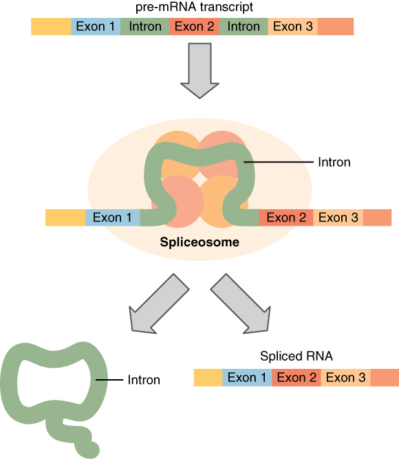
Earlier than the mRNA molecule leaves the nucleus and proceeds to protein synthesis, it’s modified in various methods. For that reason, it’s usually referred to as a pre-mRNA at this stage. For instance, your DNA, and thus complementary mRNA, incorporates lengthy areas referred to as non-coding areas that don’t code for amino acids. Their operate continues to be a thriller, however the course of referred to as splicing removes these non-coding areas from the pre-mRNA transcript (Determine 3.4.3). A spliceosome—a construction composed of assorted proteins and different molecules—attaches to the mRNA and “splices” or cuts out the non-coding areas. The eliminated phase of the transcript known as an intron. The remaining exons are pasted collectively. An exon is a phase of RNA that is still after splicing. Apparently, some introns which might be faraway from mRNA aren’t all the time non-coding. When completely different coding areas of mRNA are spliced out, completely different variations of the protein will ultimately outcome, with variations in construction and performance. This course of leads to a a lot bigger number of attainable proteins and protein features. When the mRNA transcript is prepared, it travels out of the nucleus and into the cytoplasm.
From RNA to Protein: Translation
Like translating a e-book from one language into one other, the codons on a strand of mRNA should be translated into the amino acid alphabet of proteins. Translation is the method of synthesizing a sequence of amino acids referred to as a polypeptide. Translation requires two main aids: first, a “translator,” the molecule that may conduct the interpretation, and second, a substrate on which the mRNA strand is translated into a brand new protein, just like the translator’s “desk.” Each of those necessities are fulfilled by different forms of RNA. The substrate on which translation takes place is the ribosome.
Keep in mind that a lot of a cell’s ribosomes are discovered related to the tough ER, and perform the synthesis of proteins destined for the Golgi equipment. Ribosomal RNA (rRNA) is a sort of RNA that, along with proteins, composes the construction of the ribosome. Ribosomes exist within the cytoplasm as two distinct parts, a small and a big subunit. When an mRNA molecule is able to be translated, the 2 subunits come collectively and fix to the mRNA. The ribosome offers a substrate for translation, bringing collectively and aligning the mRNA molecule with the molecular “translators” that should decipher its code.
The opposite main requirement for protein synthesis is the translator molecules that bodily “learn” the mRNA codons. Switch RNA (tRNA) is a sort of RNA that ferries the suitable corresponding amino acids to the ribosome, and attaches every new amino acid to the final, constructing the polypeptide chain one-by-one. Thus tRNA transfers particular amino acids from the cytoplasm to a rising polypeptide. The tRNA molecules should be capable to acknowledge the codons on mRNA and match them with the proper amino acid. The tRNA is modified for this operate. On one finish of its construction is a binding web site for a particular amino acid. On the opposite finish is a base sequence that matches the codon specifying its explicit amino acid. This sequence of three bases on the tRNA molecule known as an anticodon. For instance, a tRNA accountable for shuttling the amino acid glycine incorporates a binding web site for glycine on one finish. On the opposite finish it incorporates an anticodon that enhances the glycine codon (GGA is a codon for glycine, and so the tRNAs anticodon would learn CCU). Geared up with its explicit cargo and matching anticodon, a tRNA molecule can learn its acknowledged mRNA codon and produce the corresponding amino acid to the rising chain (Determine 3.4.4).
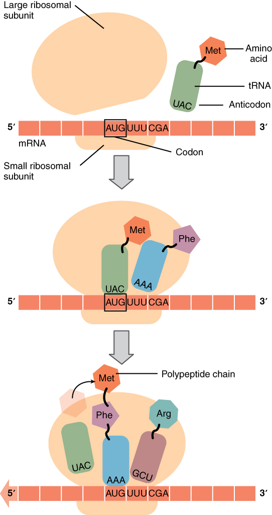
Very like the processes of DNA replication and transcription, translation consists of three primary levels: initiation, elongation, and termination. Initiation takes place with the binding of a ribosome to an mRNA transcript. The elongation stage entails the popularity of a tRNA anticodon with the subsequent mRNA codon within the sequence. As soon as the anticodon and codon sequences are sure (bear in mind, they’re complementary base pairs), the tRNA presents its amino acid cargo and the rising polypeptide strand is connected to this subsequent amino acid. This attachment takes place with the help of varied enzymes and requires power. The tRNA molecule then releases the mRNA strand, the mRNA strand shifts one codon over within the ribosome, and the subsequent acceptable tRNA arrives with its matching anticodon. This course of continues till the ultimate codon on the mRNA is reached which offers a “cease” message that alerts termination of translation and triggers the discharge of the entire, newly synthesized protein. Thus, a gene throughout the DNA molecule is transcribed into mRNA, which is then translated right into a protein product (Determine 3.4.5).
Generally, an mRNA transcription can be translated concurrently by a number of adjoining ribosomes. This will increase the effectivity of protein synthesis. A single ribosome may translate an mRNA molecule in roughly one minute; so a number of ribosomes aboard a single transcript might produce a number of instances the variety of the identical protein in the identical minute. A polyribosome is a string of ribosomes translating a single mRNA strand.
Chapter Assessment
DNA shops the knowledge needed for instructing the cell to carry out all of its features. Cells use the genetic code saved inside DNA to construct proteins, which in the end determines the construction and performance of the cell. This genetic code lies within the explicit sequence of nucleotides that make up every gene alongside the DNA molecule. To “learn” this code, the cell should carry out two sequential steps. In step one, transcription, the DNA code is transformed right into a RNA code. A molecule of messenger RNA that’s complementary to a particular gene is synthesized in a course of much like DNA replication. The molecule of mRNA offers the code to synthesize a protein. Within the technique of translation, the mRNA attaches to a ribosome. Subsequent, tRNA molecules shuttle the suitable amino acids to the ribosome, one-by-one, coded by sequential triplet codons on the mRNA, till the protein is totally synthesized. When accomplished, the mRNA detaches from the ribosome, and the protein is launched. Sometimes, a number of ribosomes connect to a single mRNA molecule without delay such that a number of proteins could be manufactured from the mRNA concurrently.
3.5 Cell Development and Division
Studying Targets
Primary Goal
- Listing the levels of the cell cycle so as, together with the steps of cell division in somatic cells
By the top of this part, it is possible for you to to:
- Describe the levels of the cell cycle
- Describe the levels of mitosis and cytokinesis, so as
- Focus on how the cell cycle is regulated
- Clarify the implications of shedding management over the cell cycle
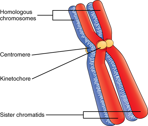
Up to now on this chapter, you’ve learn quite a few instances of the significance and prevalence of cell division. Whereas there are a couple of cells within the physique that don’t bear cell division (comparable to gametes, pink blood cells, most neurons, and a few muscle cells), most somatic cells divide frequently. A somatic cell is a common time period for a physique cell, and all human cells, aside from the cells that produce eggs and sperm (that are known as germ cells). Somatic cells comprise two copies of every of their chromosomes (one copy acquired from every father or mother). A homologous pair of chromosomes are the 2 copies of a single chromosome present in every somatic cell. The human is a diploid organism, having 23 homologous pairs of chromosomes in every of the somatic cells. The situation of getting pairs of chromosomes is named diploidy.
Cells within the physique substitute themselves over the lifetime of an individual. For instance, the cells lining the gastrointestinal tract should be continuously changed when consistently “worn off” by the motion of meals by way of the intestine. However what triggers a cell to divide, and the way does it put together for and full cell division? The cell cycle is the sequence of occasions within the lifetime of the cell from the second it’s created on the finish of a earlier cycle of cell division till it then divides itself, producing two new cells.
The Cell Cycle
One “flip” or cycle of the cell cycle consists of three common phases: interphase, adopted by mitosis and cytokinesis. Interphase is the interval of the cell cycle throughout which the cell will not be dividing. Nearly all of cells are in interphase more often than not. Mitosis is the division of genetic materials, throughout which the cell nucleus breaks down and two new, totally useful, nuclei are shaped. Cytokinesis divides the cytoplasm into two distinctive cells.
Interphase
A cell grows and carries out all regular metabolic features and processes in a interval referred to as G1 (Determine 3.5.1). G1 part (hole 1 part) is the primary hole, or development part within the cell cycle. For cells that may divide once more, G1 is adopted by replication of the DNA, in the course of the S part. The S part (synthesis part) is the interval throughout which a cell replicates its DNA.
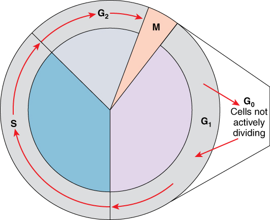
After the synthesis part, the cell proceeds by way of the G2 part. The G2 part is a second hole part, throughout which the cell continues to develop and makes the mandatory preparations for mitosis. Between G1, S, and G2 phases, cells will fluctuate essentially the most of their period of the G1 part. It’s right here {that a} cell may spend a few hours, or many days. The S part sometimes lasts between 8-10 hours and the G2 part roughly 5 hours. In distinction to those phases, the G0 part is a resting part of the cell cycle. Cells which have briefly stopped dividing and are resting (a typical situation) and cells which have completely ceased dividing (like nerve cells) are mentioned to be in G0.
The Construction of Chromosomes
Billions of cells within the human physique divide on daily basis. Through the synthesis part (S, for DNA synthesis) of interphase, the quantity of DNA throughout the cell exactly doubles. Subsequently, after DNA replication, however earlier than cell division, every cell really incorporates two copies of every chromosome. Every copy of the chromosome is known as a sister chromatid and is bodily sure to the opposite copy. The centromere is the construction that attaches one sister chromatid to a different. Since a human cell has 46 chromosomes, throughout this part, there are 92 chromatids (46 × 2) within the cell. Be sure to not confuse the idea of a pair of chromatids (one chromosome and its precise copy connected throughout mitosis) and a homologous pair of chromosomes (two paired chromosomes which had been inherited individually, one from every father or mother) (Determine 3.52).
Mitosis and Cytokinesis
The mitotic part of the cell sometimes takes between 1 and a couple of hours. Throughout this part, a cell undergoes two main processes. First, it completes mitosis, throughout which the contents of the nucleus are equitably pulled aside and distributed between its two halves. Cytokinesis then happens, dividing the cytoplasm and cell physique into two new cells. Mitosis is split into 4 main levels that happen after interphase (Determine 3.5.3) and within the following order: prophase, metaphase, anaphase, and telophase. The method is then adopted by cytokinesis.
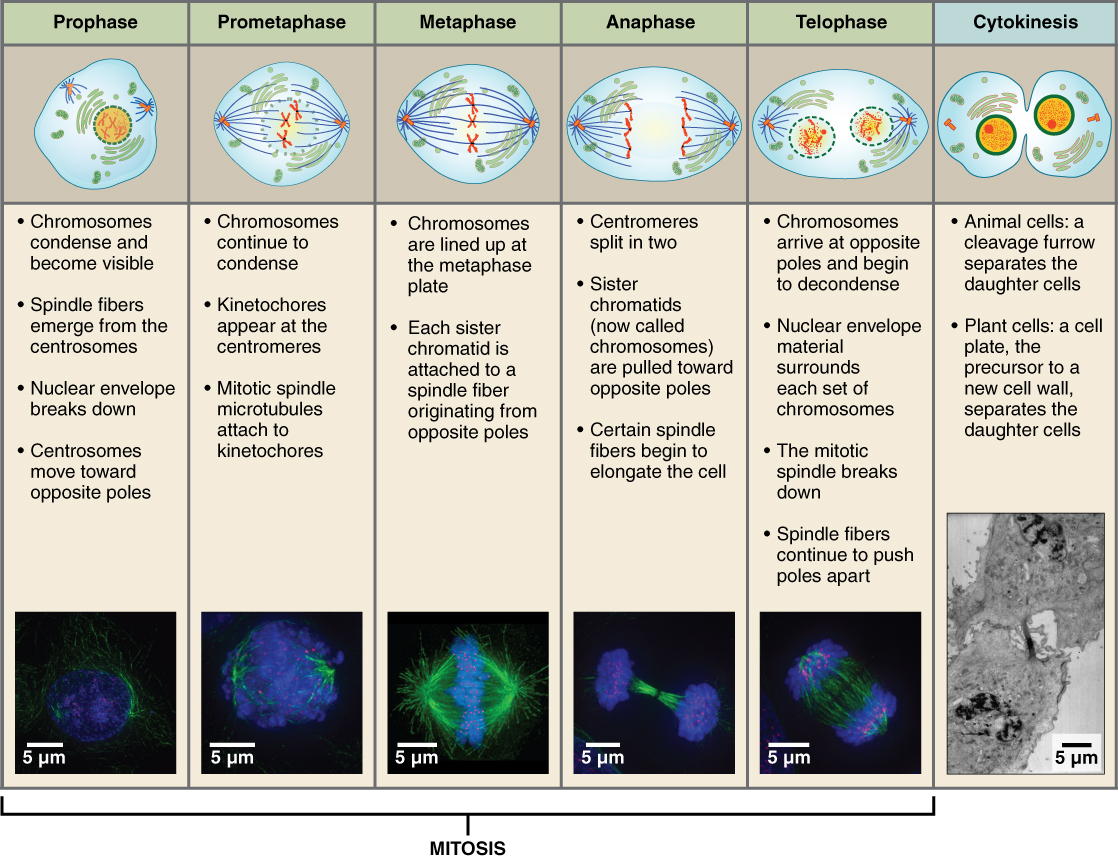
Prophase is the primary part of mitosis, throughout which the loosely packed chromatin coils and condenses into seen chromosomes. Throughout prophase, every chromosome turns into seen with its equivalent companion connected, forming the acquainted X-shape of sister chromatids. The nucleolus disappears early throughout this part, and the nuclear envelope additionally disintegrates.
A serious prevalence throughout prophase issues an important construction that incorporates the origin web site for microtubule development. Recall the mobile buildings referred to as centrioles that function origin factors from which microtubules lengthen. These tiny buildings additionally play an important function throughout mitosis. A centrosome is a pair of centrioles collectively. The cell incorporates two centrosomes side-by-side, which start to maneuver aside throughout prophase. Because the centrosomes migrate to 2 completely different sides of the cell, microtubules start to increase from every like lengthy fingers from two fingers extending towards one another. The mitotic spindle is the construction composed of the centrosomes and their rising microtubules.
Close to the top of prophase there’s an invasion of the nuclear space by microtubules from the mitotic spindle. The nuclear membrane has disintegrated, and the microtubules connect themselves to the centromeres that adjoin pairs of sister chromatids. The kinetochore is a protein construction on the centromere that’s the level of attachment between the mitotic spindle and the sister chromatids. This stage is known as late prophase or “prometaphase” to point the transition between prophase and metaphase.
Metaphase is the second stage of mitosis. Throughout this stage, the sister chromatids, with their connected microtubules, line up alongside a linear airplane in the midst of the cell. A metaphase plate kinds between the centrosomes that at the moment are situated at both finish of the cell. The metaphase plate is the title for the airplane by way of the middle of the spindle on which the sister chromatids are positioned. The microtubules at the moment are poised to drag aside the sister chromatids and produce one from every pair to every aspect of the cell.
Anaphase is the third stage of mitosis. Anaphase takes place over a couple of minutes, when the pairs of sister chromatids are separated from each other, forming particular person chromosomes as soon as once more. These chromosomes are pulled to reverse ends of the cell by their kinetochores, because the microtubules shorten. Every finish of the cell receives one companion from every pair of sister chromatids, guaranteeing that the 2 new daughter cells will comprise equivalent genetic materials.
Telophase is the ultimate stage of mitosis. Telophase is characterised by the formation of two new daughter nuclei at both finish of the dividing cell. These newly shaped nuclei encompass the genetic materials, which uncoils in such a means that the chromosomes return to loosely packed chromatin. Nucleoli additionally reappear throughout the new nuclei, and the mitotic spindle breaks aside, every new cell receiving its personal complement of DNA, organelles, membranes, and centrioles. At this level, the cell is already starting to separate in half as cytokinesis begins.
The cleavage furrow is a contractile band made up of microfilaments that kinds across the midline of the cell throughout cytokinesis. (Recall that microfilaments encompass actin). This contractile band squeezes the 2 cells aside till they lastly separate. Two new cells at the moment are shaped. Certainly one of these cells (the “stem cell”) enters its personal cell cycle; capable of develop and divide once more at some future time. The opposite cell transforms into the useful cell of the tissue, sometimes changing an “previous” cell there.
Think about a cell that accomplished mitosis however by no means underwent cytokinesis. In some instances, a cell could divide its genetic materials and develop in dimension, however fail to bear cytokinesis. This leads to bigger cells with multiple nucleus. Often that is an undesirable aberration and is usually a signal of cancerous cells.
Cell Cycle Management
A really elaborate and exact system of regulation controls direct the best way cells proceed from one part to the subsequent within the cell cycle and start mitosis. The management system entails molecules throughout the cell in addition to exterior triggers. These inner and exterior management triggers present “cease” and “advance” alerts for the cell. Exact regulation of the cell cycle is important for sustaining the well being of an organism, and lack of cell cycle management can result in most cancers.
Mechanisms of Cell Cycle Management
Because the cell proceeds by way of its cycle, every part entails sure processes that should be accomplished earlier than the cell ought to advance to the subsequent part. A checkpoint is a degree within the cell cycle at which the cycle could be signaled to maneuver ahead or stopped. At every of those checkpoints, completely different forms of molecules present the cease or go alerts, relying on sure circumstances throughout the cell. A cyclin is likely one of the main lessons of cell cycle management molecules (Determine 3.5.4). A cyclin-dependent kinase (CDK) is considered one of a bunch of molecules that work along with cyclins to find out development previous cell checkpoints. By interacting with many extra molecules, these triggers push the cell cycle ahead (until prevented from doing so by “cease” alerts, if for some purpose the cell will not be prepared). On the G1 checkpoint, the cell should be prepared for DNA synthesis to happen. On the G2 checkpoint the cell should be totally ready for mitosis. Even throughout mitosis, an important cease and go checkpoint in metaphase ensures that the cell is totally ready to finish cell division. The metaphase checkpoint ensures that every one sister chromatids are correctly connected to their respective microtubules and lined up on the metaphase plate earlier than the sign is given to separate them throughout anaphase.
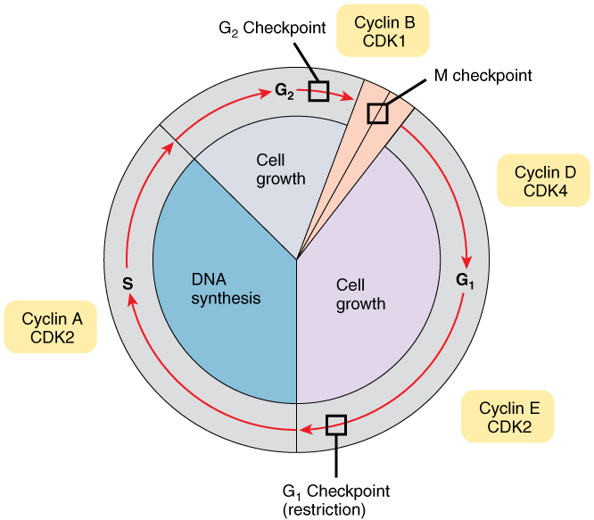
The Cell Cycle Out of Management: Implications
Most individuals perceive that most cancers or tumors are attributable to irregular cells that multiply repeatedly. If the irregular cells proceed to divide unstopped, they will injury the tissues round them, unfold to different elements of the physique, and ultimately lead to dying. In wholesome cells, the tight regulation mechanisms of the cell cycle forestall this from occurring, whereas failures of the cell cycle management could cause undesirable and extreme cell division. Failures of management could also be attributable to inherited genetic abnormalities that compromise the operate of sure “cease” and “go” alerts. Environmental insult that damages DNA may trigger dysfunction in these alerts. Usually, a mix of each genetic predisposition and environmental components result in most cancers.
The method of a cell escaping its regular management system and turning into cancerous may very well occur all through the physique fairly continuously. Luckily, sure cells of the immune system are able to recognizing cells which have turn into cancerous and destroying them. Nonetheless, in sure instances the cancerous cells stay undetected and proceed to proliferate. If the ensuing tumor doesn’t pose a menace to surrounding tissues, it’s mentioned to be benign and might normally be simply eliminated. If able to injury, the tumor is taken into account malignant and the affected person is recognized with most cancers.
Homeostatic Imbalances: Most cancers Arises from Homeostatic Imbalances
Most cancers is an especially complicated situation, able to arising from all kinds of genetic and environmental causes. Sometimes, mutations or aberrations in a cell’s DNA that compromise regular cell cycle management programs result in cancerous tumors. Cell cycle management is an instance of a homeostatic mechanism that maintains correct cell operate and well being. Whereas progressing by way of the phases of the cell cycle, a big number of intracellular molecules present cease and go alerts to control motion ahead to the subsequent part. These alerts are maintained in an intricate steadiness in order that the cell solely proceeds to the subsequent part when it’s prepared. This homeostatic management of the cell cycle could be considered like a automobile’s cruise management. Cruise management will frequently apply simply the correct quantity of acceleration to take care of a desired pace, until the motive force hits the brakes, during which case the automobile will decelerate. Equally, the cell consists of molecular messengers, comparable to cyclins, that push the cell ahead in its cycle.
Along with cyclins, a category of proteins which might be encoded by genes referred to as proto-oncogenes present vital alerts that regulate the cell cycle and transfer it ahead. Examples of proto-oncogene merchandise embrace cell-surface receptors for development components, or cell-signaling molecules, two lessons of molecules that may promote DNA replication and cell division. In distinction, a second class of genes generally known as tumor suppressor genes sends cease alerts throughout a cell cycle. For instance, sure protein merchandise of tumor suppressor genes sign potential issues with the DNA and thus cease the cell from dividing, whereas different proteins sign the cell to die whether it is broken past restore. Some tumor suppressor proteins additionally sign a ample surrounding mobile density, which signifies that the cell needn’t presently divide. The latter operate is uniquely vital in stopping tumor development. Regular cells exhibit a phenomenon referred to as “contact inhibition”, thus, intensive mobile contact with neighboring cells causes a sign that stops additional cell division.
These two contrasting lessons of genes, proto-oncogenes and tumor suppressor genes, are just like the accelerator and brake pedal of the cell’s personal “cruise management system,” respectively. Below regular circumstances, these cease and go alerts are maintained in a homeostatic steadiness. Typically talking, there are two ways in which the cell’s cruise management can lose management: a malfunctioning (overactive) accelerator, or a malfunctioning (underactive) brake. When compromised by way of a mutation, or in any other case altered, proto-oncogenes could be transformed to oncogenes, which produce oncoproteins that push a cell ahead in its cycle and stimulate cell division even when it’s undesirable to take action. For instance, a cell that needs to be programmed to self-destruct (a course of referred to as apoptosis) resulting from intensive DNA injury may as a substitute be triggered to proliferate by an oncoprotein. Alternatively, a dysfunctional tumor suppressor gene could fail to supply the cell with a needed cease sign, additionally leading to undesirable cell division and proliferation.
A fragile homeostatic steadiness between the various proto-oncogenes and tumor suppressor genes delicately controls the cell cycle and ensures that solely wholesome cells replicate. Subsequently, a disruption of this homeostatic steadiness could cause aberrant cell division and cancerous growths.
Chapter Assessment
The lifetime of cell consists of levels that make up the cell cycle. After a cell is born, it passes by way of an interphase earlier than it is able to replicate itself and produce daughter cells. This interphase consists of two hole phases (G1 and G2), in addition to an S part, throughout which its DNA is replicated in preparation for cell division. The cell cycle is underneath exact regulation by chemical messengers each inside and outdoors the cell that present “cease” and “go” alerts for motion from one part to the subsequent. Failures of those alerts can lead to cells that proceed to divide uncontrollably, which may result in most cancers.
As soon as a cell has accomplished interphase and is prepared for cell division, it proceeds by way of 4 separate levels of mitosis (prophase, metaphase, anaphase, and telophase). Telophase is adopted by the division of the cytoplasm (cytokinesis), which generates two daughter cells. This course of takes place in all usually dividing cells of the physique aside from the germ cells that produce eggs and sperm.
3.6 Mobile Differentiation
Studying Targets
Primary Goal:
- Focus on how a cell differentiates and turns into extra specialised
By the top of this part, it is possible for you to to:
- Focus on how the generalized cells of a creating embryo, or the stem cells of an grownup organism, turn into differentiated into specialised cells
- Distinguish between the classes of stem cells
How does a fancy organism comparable to a human develop from a single cell—a fertilized egg—into the huge array of cell sorts comparable to nerve cells, muscle cells, and epithelial cells that characterize the grownup? All through improvement and maturity, the method of mobile differentiation leads cells to imagine their remaining morphology and physiology. Differentiation is the method by which unspecialized cells turn into specialised to hold out distinct features.
Stem Cells
A stem cell is an unspecialized cell that may divide with out restrict as wanted and might, underneath particular circumstances, differentiate into specialised cells. Stem cells are divided into a number of classes in accordance with their potential to distinguish.
The primary embryonic cells that come up from the division of the zygote are the last word stem cells; these stems cells are described as totipotent as a result of they’ve the potential to distinguish into any of the cells wanted to allow an organism to develop and develop.
The embryonic cells that develop from totipotent stem cells and are precursors to the elemental tissue layers of the embryo are categorised as pluripotent. A pluripotent stem cell is one which has the potential to distinguish into any kind of human tissue however can’t assist the total improvement of an organism. These cells then turn into barely extra specialised, and are known as multipotent cells.
A multipotent stem cell has the potential to distinguish into various kinds of cells inside a given cell lineage or small variety of lineages, comparable to a pink blood cell or white blood cell.
Lastly, multipotent cells can turn into additional specialised oligopotent cells. An oligopotent stem cell is restricted to turning into one of some completely different cell sorts. In distinction, a unipotent cell is totally specialised and might solely reproduce to generate extra of its personal particular cell kind.
Stem cells are distinctive in that they will additionally frequently divide and regenerate new stem cells as a substitute of additional specializing. There are completely different stem cells current at completely different levels of a human’s life. They embrace the embryonic stem cells of the embryo, fetal stem cells of the fetus, and grownup stem cells within the grownup. One kind of grownup stem cell is the epithelial stem cell, which supplies rise to the keratinocytes within the a number of layers of epithelial cells within the dermis of pores and skin. Grownup bone marrow has three distinct forms of stem cells: hematopoietic stem cells (which give rise to pink blood cells, white blood cells, and platelets), endothelial stem cells (which give rise to the endothelial cell sorts that line blood and lymph vessels), and mesenchymal stem cells (which give rise to the various kinds of muscle cells).
The method of hematopoiesis entails the differentiation of multipotent cells into blood and immune cells. The multipotent hematopoietic stem cells give rise to many various cell sorts, together with the cells of the immune system and pink blood cells.
Differentiation
When a cell differentiates (turns into extra specialised), it might undertake main adjustments in its dimension, form, metabolic exercise, and general operate. Since all cells within the physique, starting with the fertilized egg, comprise the identical DNA, how do the completely different cell sorts come to be so completely different? The reply is analogous to a film script. The completely different actors in a film all learn from the identical script, nonetheless, they’re every solely studying their very own a part of the script. Equally, all cells comprise the identical full complement of DNA, however every kind of cell solely “reads” the parts of DNA which might be related to its personal operate. In biology, that is known as the distinctive genetic expression of every cell.
To ensure that a cell to distinguish into its specialised type and performance, it want solely manipulate these genes (and thus these proteins) that can be expressed, and never these that may stay silent. The first mechanism by which genes are turned “on” or “off” is thru transcription components.
Whereas every physique cell incorporates the organism’s total genome, completely different cells regulate gene expression with the usage of varied transcription components. Transcription components are proteins that have an effect on the binding of RNA polymerase to a selected gene on the DNA molecule.
On a regular basis Connection: Stem Cell Analysis
Stem cell analysis goals to search out methods to make use of stem cells to regenerate and restore mobile injury. Over time, most grownup cells bear the damage and tear of getting older and lose their potential to divide and restore themselves. Stem cells don’t show a selected morphology or operate. Grownup stem cells, which exist as a small subset of cells in most tissues, hold dividing and might differentiate into various specialised cells usually shaped by that tissue. These cells allow the physique to resume and restore physique tissues.
The mechanisms that induce a non-differentiated cell to turn into a specialised cell are poorly understood. In a laboratory setting, it’s attainable to induce stem cells to distinguish into specialised cells by altering the bodily and chemical circumstances of development. A number of sources of stem cells are used experimentally and are categorised in accordance with their origin and potential for differentiation. Human embryonic stem cells (hESCs) are extracted from embryos and are pluripotent. The grownup stem cells which might be current in lots of organs and differentiated tissues, comparable to bone marrow and pores and skin, are multipotent, being restricted in differentiation to the forms of cells present in these tissues. The stem cells remoted from umbilical wire blood are additionally multipotent, as are cells from deciduous tooth (child tooth). Researchers have not too long ago developed induced pluripotent stem cells (iPSCs) from mouse and human grownup stem cells. These cells are genetically reprogrammed multipotent grownup cells that operate like embryonic stem cells; they’re able to producing cells attribute of all three germ layers.
Due to their capability to divide and differentiate into specialised cells, stem cells supply a possible remedy for illnesses comparable to diabetes and coronary heart illness (Determine 3.6.1). Cell-based remedy refers to remedy during which stem cells induced to distinguish in a development dish are injected right into a affected person to restore broken or destroyed cells or tissues. Many obstacles should be overcome for the applying of cell-based remedy. Though embryonic stem cells have a virtually limitless vary of differentiation potential, they’re seen as overseas by the affected person’s immune system and should set off rejection. Additionally, the destruction of embryos to isolate embryonic stem cells raises appreciable moral and authorized questions.
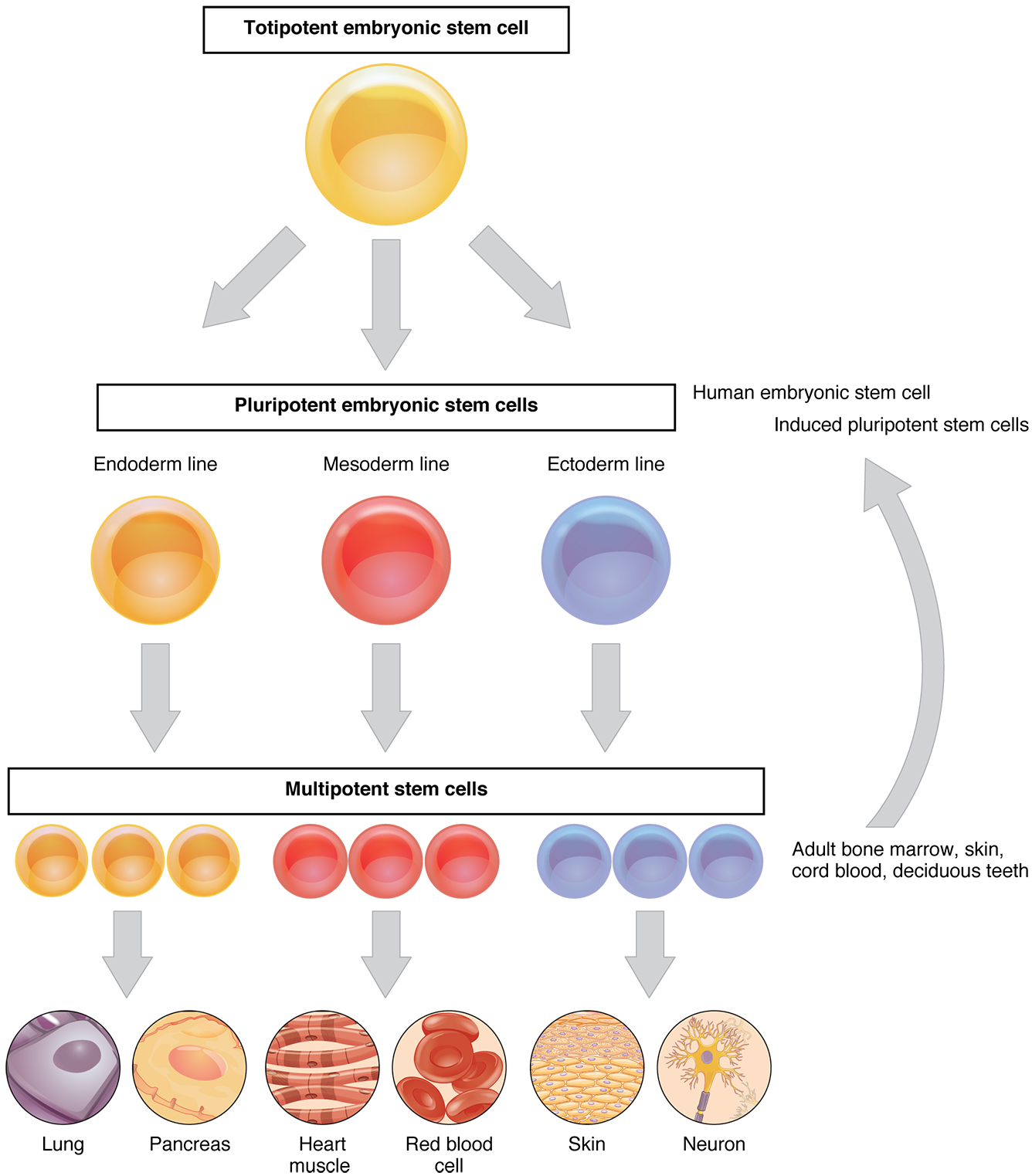
In distinction, grownup stem cells remoted from a affected person aren’t seen as overseas by the physique, however they’ve a restricted vary of differentiation. Some people financial institution the wire blood or deciduous tooth of their baby, storing away these sources of stem cells for future use, ought to their baby want it. Induced pluripotent stem cells are thought of a promising advance within the subject as a result of utilizing them avoids the authorized, moral, and immunological pitfalls of embryonic stem cells.
Chapter Assessment
One of many main areas of analysis in biology is of how cells specialize to imagine their distinctive buildings and features, since all cells basically originate from a single fertilized egg. Cell differentiation is the method of cells turning into specialised as their physique develops. A stem cell is an unspecialized cell that may divide with out restrict as wanted and might, underneath particular circumstances, differentiate into specialised cells. Stem cells are divided into a number of classes in accordance with their potential to distinguish. Whereas all somatic cells comprise the very same genome, completely different cell sorts solely specific a few of these genes at any given time. These variations in gene expression in the end dictate a cell’s distinctive morphological and physiological traits. The first mechanism that determines which genes can be expressed and which of them won’t, is thru the usage of completely different transcription issue proteins, which bind to DNA and promote or hinder the transcription of various genes. By the motion of those transcription components, cells specialize into considered one of a whole bunch of various cell sorts within the human physique.